 뒤로
뒤로Microscopy, Histology, and Tissue Types: Foundations for Anatomy & Physiology
스터디 가이드 - 스마트 노트
 자료에 맞춘 맞춤형 노트, 핵심 정의, 예시, 맥락을 확장해 제공합니다.
Microscopy in Anatomy & Physiology
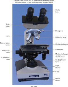
Microscope Anatomy and Function
Microscopy is essential for studying cells and tissues in anatomy and physiology. The compound light microscope is the primary tool used to observe thin sections of biological specimens.
Ocular lens: The eyepiece through which you view the specimen.
Objective lens: Provides varying levels of magnification (e.g., 4x, 10x, 43x).
Stage: Platform where the slide is placed.
Coarse-focus and fine-focus knobs: Adjust the clarity of the image.
Condenser and iris diaphragm: Control light intensity and focus.
Base and arm: Provide structural support.
Microscope Slide Preparation
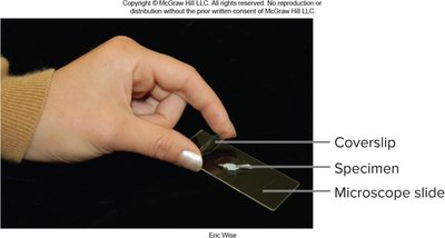
Specimens are placed on glass slides and covered with a coverslip to protect the sample and improve viewing.
Microscope slide: Flat glass platform for specimen.
Coverslip: Thin glass piece placed over the specimen.
Magnification and Field of View
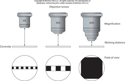
The total magnification is calculated by multiplying the ocular lens magnification by the objective lens magnification. Higher magnification reduces the field of view and working distance.
Formula:
Field of view: The visible area under the microscope decreases as magnification increases.
Working distance: The space between the objective lens and the specimen decreases with higher magnification.
Histology: The Study of Tissues
The Tissue Level of Organization
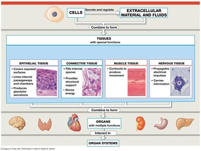
Tissues are collections of specialized cells and their products that perform specific functions. Histology is the study of tissue structure, which is fundamental for understanding organ function.
Organs: Composed of two or more tissue types.
Histology: Examines tissue structure and function.
Four Basic Types of Tissue
The human body contains four primary tissue types, each with distinct functions:
Epithelial tissue: Covers surfaces, lines passageways, forms glands.
Connective tissue: Supports, connects, transports, and stores energy.
Muscle tissue: Specialized for contraction and movement.
Nervous tissue: Conducts electrical impulses and processes information.
Epithelial Tissue
Structure and Function
Epithelial tissue includes layers of cells covering internal and external surfaces and forms glands. It serves several key functions:
Physical protection: Shields surfaces from abrasion and pathogens.
Control permeability: Regulates entry and exit of substances.
Provide sensation: Contains sensory receptors for various stimuli.
Produce secretions: Gland cells release substances for lubrication, protection, or communication.
Characteristics of Epithelial Tissue
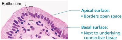
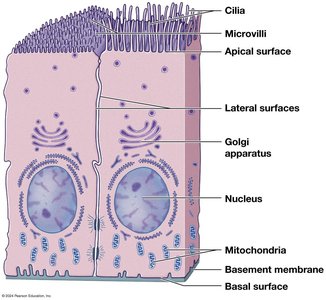
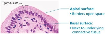
Polarity: Distinct apical (exposed) and basal (attached) surfaces.
Cellularity: Cells are tightly bound by junctions.
Attachment: Base is anchored to a basement membrane.
Avascularity: Lacks blood vessels; nutrients diffuse from underlying connective tissue.
Regeneration: Rapid replacement by stem cell division.
Classification of Epithelia
Epithelia are classified by cell shape and number of layers:
Shapes: Squamous (flat), cuboidal (boxy), columnar (tall).
Layers: Simple (one layer), stratified (multiple layers).
Squamous Epithelium
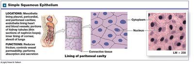
Simple squamous: Absorption and diffusion; lines alveoli, blood vessels.
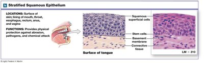
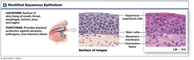
Stratified squamous: Protection; found in skin, mouth, esophagus.
Keratinized: Epidermis, gums, hard palate.
Non-keratinized: Mouth, esophagus, vagina.
Cuboidal Epithelium
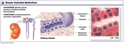
Simple cuboidal: Secretion and absorption; glands, kidney tubules.
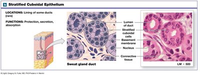
Stratified cuboidal: Rare; ducts of sweat and mammary glands.
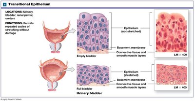
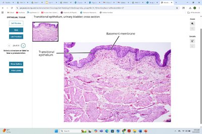
Transitional Epithelium
Appearance: Cuboidal when relaxed, squamous when stretched.
Location: Urinary bladder; tolerates stretching.
Columnar Epithelium
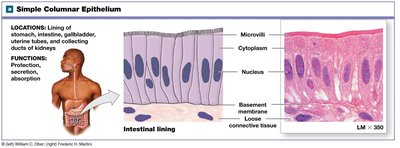
Simple columnar: Absorption and secretion; stomach, intestines.
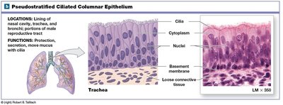
Pseudostratified columnar: Appears layered, has cilia; nasal cavity, trachea.
Stratified columnar: Rare; protection in pharynx, anus, urethra.
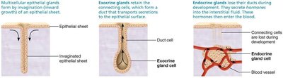
Glandular Epithelia
Endocrine glands: Release hormones into bloodstream; no ducts.
Exocrine glands: Discharge secretions onto surfaces via ducts.
Connective Tissue
Overview and Functions
Connective tissue is the most abundant tissue type, providing structural support, transport, protection, energy storage, and defense.
Classes: Connective tissue proper, cartilage, bone, blood.
Functions: Framework, transport, protection, support, energy storage, defense.
Common Characteristics
Origin: Derived from mesenchyme.
Vascularity: Varies from avascular (cartilage) to highly vascular (bone).
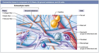
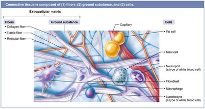
Extracellular matrix: Composed of ground substance and fibers; supports cells.
Structural Elements
Ground substance: Glycoproteins and polysaccharides; syrupy or gelatinous.
Fibers: Collagen (strength), elastic (stretch), reticular (support).
Cells: "Blast" (immature, matrix-secreting), "cyte" (mature, maintenance), fat cells, white blood cells, mast cells, macrophages.
Summary Table: Tissue Types and Functions
Tissue Type | Main Function | Example Location |
|---|---|---|
Epithelial | Protection, secretion, absorption | Skin, lining of organs |
Connective | Support, transport, energy storage | Bone, blood, fat |
Muscle | Contraction, movement | Skeletal muscles, heart |
Nervous | Electrical signaling, information processing | Brain, nerves |
Key Points for Exam Preparation
Understand microscope anatomy and slide preparation.
Know the four basic tissue types and their functions.
Be able to classify epithelial tissues by shape and layers.
Recognize the structural elements of connective tissue.
Apply the formula for total magnification in microscopy.
Additional info: Academic context was added to clarify tissue functions, microscope operation, and classification schemes. Images were included only when directly relevant to the explanation of the paragraph.
