 뒤로
뒤로Module 1: Diffusion, Osmosis, Cell Structure, Cell Cycle, and Anatomical Terminology
스터디 가이드 - 스마트 노트
 자료에 맞춘 맞춤형 노트, 핵심 정의, 예시, 맥락을 확장해 제공합니다.
Diffusion and Osmosis
Diffusion
Diffusion is the passive movement of molecules from an area of higher concentration to an area of lower concentration. This process is fundamental for the transport of substances within biological systems.
Occurs in liquids and gases, but not in solids due to fixed molecular positions.
Permeability: A membrane must be permeable for diffusion to occur; impermeable membranes prevent diffusion.
Temperature effect: Higher temperatures increase molecular vibration and diffusion rate.
Osmosis
Osmosis is the movement of water molecules across a selectively permeable membrane from an area of higher water concentration (lower solute concentration) to an area of lower water concentration (higher solute concentration).
Essential for maintaining cell volume and fluid balance.
Types of Solutions and Effects on Cells
Isotonic Solution: Equal solute concentration inside and outside the cell; no net water movement.
Hypertonic Solution: Higher solute concentration outside the cell; water moves out, causing the cell to shrink (crenation).
Hypotonic Solution: Lower solute concentration outside the cell; water moves in, causing the cell to swell and possibly burst (lysis).
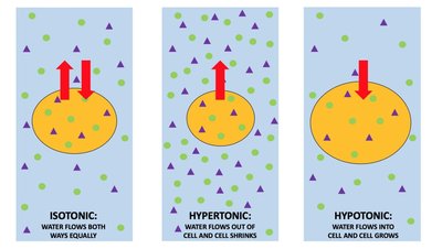
Example: Red blood cells in different solutions demonstrate these effects: swelling in hypotonic, shrinking in hypertonic, and maintaining shape in isotonic solutions.
Cell Structure and Function
Plasma Membrane
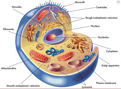
The plasma membrane is a thin, flexible barrier that surrounds the cell, controlling the movement of substances in and out.
Acts as a boundary between the cell's internal environment and the external world.
Nucleus and Nucleolus
Nucleus: The control center of the cell, containing genetic material (DNA) and directing cellular activities such as growth and reproduction.
Nucleolus: Located inside the nucleus, responsible for ribosome production.
Chromosomes
Structure: DNA tightly packed with histone proteins.
Function: Carry genes that encode instructions for protein synthesis.
Human cells: 46 chromosomes (23 pairs).
Cytoplasmic Organelles
Ribosomes: Sites of protein synthesis, composed of rRNA and proteins.
Rough Endoplasmic Reticulum (ER): Studded with ribosomes; synthesizes and processes proteins.
Smooth Endoplasmic Reticulum (SER): Involved in lipid synthesis, detoxification, and storage.
Golgi Apparatus: Modifies, sorts, and packages proteins and lipids for delivery.
Mitochondria: Powerhouse of the cell; site of ATP production via cellular respiration.
Centrioles: Organize microtubules and play a role in cell division.
Microvilli: Increase surface area for absorption.
Example: The mitochondria convert glucose into ATP, providing energy for cellular processes.
Cell Cycle
Interphase
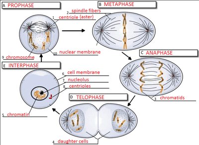
Interphase is the period of cell growth and DNA replication, preparing the cell for division.
G1, S, G2 phases: Cell grows, duplicates organelles, and replicates DNA.
Nuclear envelope is intact, and DNA is in chromatin form.
Mitosis (M Phase)
Mitosis is the process by which a cell divides to produce two genetically identical daughter cells.
Prophase: Chromatin condenses into visible chromosomes; nuclear envelope breaks down; spindle fibers form.
Metaphase: Chromosomes align at the cell's equator.
Anaphase: Sister chromatids separate and move to opposite poles.
Telophase: Nuclear envelopes reform; chromosomes decondense; cytokinesis divides the cytoplasm.
Example: Skin cells undergo mitosis to replace damaged or dead cells.
Anatomical Terminology
Regional Terms
Regional terms specify distinct areas of the body, aiding in precise anatomical description.
Cephalic: Head
Facial: Face
Thoracic: Chest/ribcage
Abdominal: Abdomen/torso
Pelvic: Hips
Upper limb: Arm regions (e.g., brachial, antecubital, carpal)
Lower limb: Leg regions (e.g., femoral, crural, tarsal)
Body Positions
Prone: Lying on the stomach
Supine: Lying on the back
Directional Terms
Anterior (ventral): Front of the body
Posterior (dorsal): Back of the body
Medial: Toward the midline
Lateral: Away from the midline
Superior: Above
Inferior: Below
Proximal: Closer to the point of attachment
Distal: Farther from the point of attachment
Superficial: Closer to the surface
Deep: Further from the surface
Body Planes
Frontal (coronal): Divides body into anterior and posterior
Transverse (horizontal): Divides body into superior and inferior
Sagittal: Divides body into left and right
Mid-sagittal: Divides body into equal left and right halves
Parasagittal: Divides body into unequal left and right portions
Oblique: Slanted division
Tissues
Epithelial Tissue
Function: Lines surfaces and cavities; forms glands.
Types: Simple (one layer), stratified (multiple layers).
Shapes: Squamous (flat), cuboidal (cube-shaped), columnar (tall).
Connective Tissue
Function: Supports, binds, and protects organs.
Types: Bone (hard, calcified), cartilage (soft, cushioning), blood (cells in plasma), lymph (fluid outside blood vessels).
Tendons: Connect muscle to bone.
Ligaments: Connect bone to bone.
Muscle Tissue
Cardiac: Striated, single nucleus, involuntary, found in heart.
Skeletal: Striated, multiple nuclei, voluntary, attached to bones.
Smooth: Non-striated, spindle-shaped, involuntary, found in walls of organs.
Nervous Tissue
Neurons: Specialized for communication and coordination of body functions.
Integumentary System
Structure
Cutaneous membrane: Skin
Accessory structures: Hair, nails, sweat glands, etc.
Layers of the Skin
Epidermis: Epithelial tissue; layers include (from superficial to deep):
Stratum corneum
Stratum granulosum
Stratum spinosum
Stratum basale
Stratum lucidum (only in thick skin)
Dermis: Connective tissue; contains papillary (superficial) and reticular (deep) layers, sensory receptors (Meissner's and Pacinian corpuscles), sebaceous (oil) and sudoriferous (sweat) glands.
Hypodermis: Deepest layer; connects skin to underlying tissues.
Hair Structure
Hair follicle: Anchors hair in skin.
Hair bulb: Base of follicle; site of hair growth.
Hair papilla: Supplies nutrients to growing hair.
Arrector pili: Muscle that causes hair to stand up.
Example: The epidermis provides a barrier to pathogens, while sweat glands help regulate body temperature.
Additional info: Some tissues, like cartilage, heal slowly due to lack of blood supply, while bone heals faster because it is vascularized.
