 뒤로
뒤로Muscle and Nervous Tissues: Structure and Function
스터디 가이드 - 스마트 노트
 자료에 맞춘 맞춤형 노트, 핵심 정의, 예시, 맥락을 확장해 제공합니다.
Muscle Tissue
Overview of Muscle Tissue
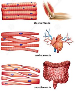
Muscle tissue is responsible for most types of movement in the human body. It is classified into three main types, each with distinct structural and functional characteristics.
Skeletal muscle tissue: Found in skeletal muscles attached to bones; responsible for voluntary movements.
Cardiac muscle tissue: Located in the walls of the heart; controls involuntary contractions that pump blood.
Smooth muscle tissue: Present mainly in the walls of hollow organs (other than the heart); regulates involuntary movements such as peristalsis.
Skeletal Muscle Tissue
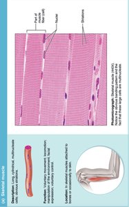
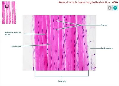
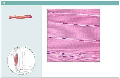
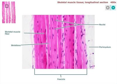
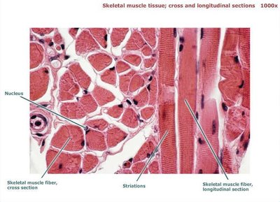
Skeletal muscle tissue consists of long, cylindrical, multinucleate cells with obvious striations. It is under voluntary control and is essential for locomotion, manipulation of the environment, and facial expression.
Description: Long, cylindrical, multinucleate cells; striated appearance.
Function: Voluntary movement; locomotion; manipulation of the environment; facial expression.
Location: Skeletal muscles attached to bones or occasionally to skin.
Key Features: Striations, multiple nuclei, muscle fibers (cells).
Cardiac Muscle Tissue
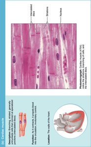
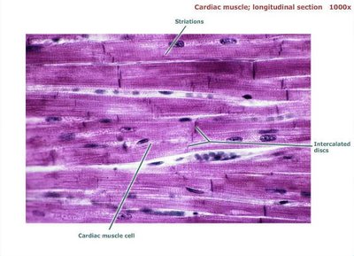
Cardiac muscle tissue is found only in the heart. It is composed of branching, striated cells that are generally uninucleate and connected by intercalated discs. Cardiac muscle is involuntary and responsible for pumping blood throughout the body.
Description: Branching, striated, generally uninucleate cells; intercalated discs present.
Function: Involuntary contraction; propels blood into circulation.
Location: Walls of the heart.
Key Features: Striations, intercalated discs, single nucleus per cell.
Smooth Muscle Tissue
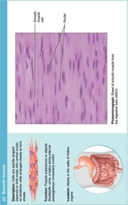
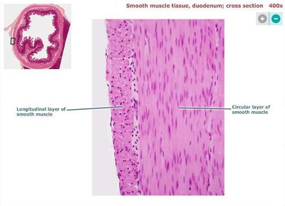
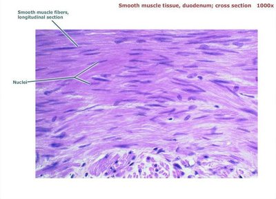
Smooth muscle tissue consists of spindle-shaped cells with a single central nucleus and no visible striations. It is found in the walls of hollow organs and is responsible for involuntary movements such as peristalsis and regulation of blood vessel diameter.
Description: Spindle-shaped cells; single central nucleus; no striations.
Function: Propels substances or objects along internal passageways; involuntary control.
Location: Walls of hollow organs (e.g., intestines, blood vessels).
Key Features: Smooth appearance, centrally located nuclei.
Nervous Tissue
Overview of Nervous Tissue
Nervous tissue is the main component of the nervous system, including the brain, spinal cord, and nerves. It regulates and controls body functions by transmitting electrical impulses.
Neurons: Specialized nerve cells that generate and conduct nerve impulses.
Neuroglia: Supporting cells that support, insulate, and protect neurons.
CNS Neuroglia: Astrocytes, Microglia, Ependymal cells, Oligodendrocytes.
PNS Neuroglia: Satellite cells, Schwann cells.
Neurons: Structure and Function
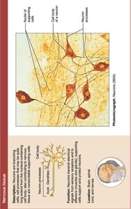
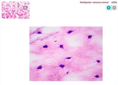
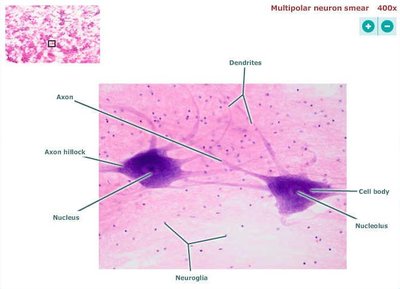
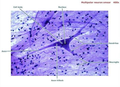
Neurons are the structural units of the nervous system. They are large, highly specialized cells that conduct impulses. Each neuron consists of a cell body (soma) and one or more processes (dendrites and axons).
Cell Body (Soma): Biosynthetic center; synthesizes proteins, membranes, and other chemicals; contains rough ER (Nissl bodies) and nucleus with nucleolus.
Dendrites: Receptive/input regions of the neuron; receive signals from other cells.
Axon: Conducting/output region; transmits impulses away from the cell body.
Axon Hillock: Region where the axon originates from the cell body.
Myelin Sheath: Insulates axons, increases impulse speed; gaps called nodes of Ranvier.
Terminal Branches: Endings of the axon; form synapses with other cells.
Neuroglia: Types and Functions
Neuroglia are supporting cells in the nervous system. They provide structural support, insulation, and protection for neurons. Different types of neuroglia are found in the central and peripheral nervous systems.
Astrocytes: Support and brace neurons; help form the blood-brain barrier.
Microglia: Act as phagocytes; remove debris and pathogens.
Ependymal Cells: Line cerebrospinal fluid-filled cavities; help circulate CSF.
Oligodendrocytes: Form myelin sheaths in the CNS.
Satellite Cells: Surround neuron cell bodies in the PNS.
Schwann Cells: Form myelin sheaths in the PNS.
Summary Table: Muscle Tissue Types
Type | Location | Control | Key Features |
|---|---|---|---|
Skeletal | Attached to bones | Voluntary | Striated, multinucleate |
Cardiac | Heart walls | Involuntary | Striated, intercalated discs, uninucleate |
Smooth | Walls of hollow organs | Involuntary | No striations, spindle-shaped, single nucleus |
Summary Table: Nervous Tissue Cell Types
Cell Type | Location | Function |
|---|---|---|
Neuron | CNS & PNS | Conducts impulses |
Astrocyte | CNS | Support, blood-brain barrier |
Microglia | CNS | Phagocytosis |
Ependymal cell | CNS | Circulate CSF |
Oligodendrocyte | CNS | Myelin sheath formation |
Satellite cell | PNS | Support neuron cell bodies |
Schwann cell | PNS | Myelin sheath formation |
Additional info: Academic context was added to expand brief points and clarify the structure and function of muscle and nervous tissues, as well as the roles of neuroglia.
