 뒤로
뒤로Neuronal Physiology and Cell Communication: Study Notes
스터디 가이드 - 스마트 노트
 자료에 맞춘 맞춤형 노트, 핵심 정의, 예시, 맥락을 확장해 제공합니다.
Neuronal Physiology
Communication Throughout the Body
The body utilizes two primary signaling systems to coordinate physiological functions: the nervous system and the endocrine system. Both systems use excitable tissues, such as nerves and muscles, which can undergo rapid changes in membrane potential to generate electrical signals.
Nervous System: Fast, short-term signaling.
Endocrine System: Moderate speed, can be long or short-term.
Excitable Tissues: Nerve and muscle cells capable of generating electrical signals.
Changes in Membrane Potential (Vm)
Membrane potential refers to the voltage difference across a cell membrane. Changes in this potential are essential for electrical signaling in neurons and muscle cells.
Polarization: Any state other than 0 mV; resting membrane potential (Vrest) is typically negative.
Depolarization: Membrane potential becomes less negative.
Repolarization: Return to resting membrane potential.
Hyperpolarization: Membrane potential becomes more negative than resting.
Electrical signals are generated by the movement of ions through membrane channels, altering the membrane's permeability.
Types of Change in Vm
Graded Potentials: Local, short-distance changes in membrane potential. The magnitude depends on the strength of the stimulus.
Action Potentials: All-or-nothing, long-distance signals that always produce the same change in potential once threshold is reached.
Graded Potentials
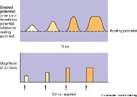
Graded potentials are local changes in membrane potential whose strength is proportional to the stimulus. They occur when gated ion channels open, allowing ions to flow and depolarize or hyperpolarize the membrane.
Greater stimulus opens more channels, resulting in a larger potential change.
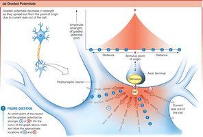
Graded potentials decrease in strength as they spread from the point of origin due to current leak out of the cell.
Spreading of Graded Potentials
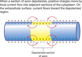
Graded potentials spread by local current flow, where positive ions move away from the depolarized region, causing adjacent areas to depolarize. This spread is bidirectional but diminishes with distance.
Action Potentials
Action potentials are rapid, large changes in membrane potential that propagate without decrement along the axon. They are initiated when graded potentials depolarize the membrane to a threshold value (typically about -50 mV).
All-or-none response: Once threshold is reached, the same amplitude is always produced.
Consist of rapid depolarization (up to +30 mV), followed by repolarization and often a brief hyperpolarization.
Membrane Permeability and Vm
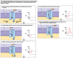
Action potentials are generated by the opening and closing of voltage-gated ion channels:
Voltage-gated Na+ channels: Have activation and inactivation gates, cycling through closed, open, and inactivated states.
Voltage-gated K+ channels: Have a single gate and open more slowly than Na+ channels.
Na+/K+ Pump
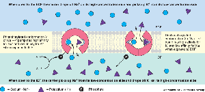
After an action potential, the Na+ and K+ concentration gradients are restored by the Na+/K+ ATPase pump, which moves Na+ out of and K+ into the cell.
Neuron Structure and Function
Neurons are the functional units of the nervous system, specialized for carrying electrical signals.
Cell Body: Control center of the neuron.
Dendrites: Receive incoming signals (graded potentials).
Axon: Conducts outgoing action potentials from the axon hillock to axon terminals.
Nerve Conduction
Action potentials are conducted along axons by two main methods:
Contiguous Conduction: Action potentials spread to adjacent regions of the axon in unmyelinated fibers.
Saltatory Conduction: In myelinated fibers, action potentials jump from node to node, increasing conduction speed.
Refractory Period
The refractory period ensures one-way propagation of action potentials and limits their frequency.
Absolute Refractory Period: No new action potential can be generated (inactivation gate closed).
Relative Refractory Period: A stronger stimulus is required to generate another action potential (some K+ channels still open).
Synapses
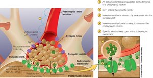
Synapses are specialized junctions where neurons communicate with other neurons, muscles, or glands. The presynaptic neuron releases neurotransmitters that bind to receptors on the postsynaptic cell, altering its membrane potential.
Synapse Structure: Includes presynaptic axon terminal, synaptic cleft, and postsynaptic membrane.
Neurotransmitter Release: Triggered by Ca2+ influx at the axon terminal, leading to exocytosis of neurotransmitter vesicles.
Postsynaptic Response: Neurotransmitter binding opens specific ion channels, generating graded responses.
Excitatory and Inhibitory Responses
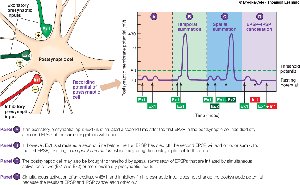
Synaptic responses can be excitatory (EPSP) or inhibitory (IPSP):
EPSP (Excitatory Postsynaptic Potential): Depolarizes the postsynaptic membrane, usually subthreshold.
IPSP (Inhibitory Postsynaptic Potential): Hyperpolarizes the postsynaptic membrane, making action potential generation less likely.
Grand Postsynaptic Potential and Summation
Multiple EPSPs and IPSPs are integrated at the postsynaptic neuron. Summation can be temporal (from one synapse in rapid succession) or spatial (from multiple synapses simultaneously). The net effect determines whether an action potential is generated.
Cell-to-Cell Communication
Physiological Signals
Cells communicate using electrical and chemical signals. Chemical signals, or extracellular chemical messengers (ECMs), are the primary means of communication and act as ligands binding to specific receptors on target cells.
Specificity, affinity, competition, and saturation are key properties of ligand-receptor interactions.
Local Communication
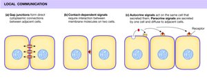
Local communication occurs through:
Gap Junctions: Direct cytoplasmic connections between adjacent cells.
Contact-Dependent Signals: Require direct membrane contact.
Paracrine and Autocrine Signals: ECMs diffuse through extracellular fluid to act on nearby or the same cell.
Long-Distance Communication
Long-distance communication uses both chemical and electrical signals:
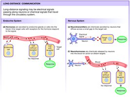
Hormones: Secreted by endocrine glands, travel in the blood to distant targets.
Neurotransmitters: Released by neurons, act on nearby cells.
Neurohormones: Released by neurons into the blood, act at distant sites.
Extracellular Chemical Messengers (ECMs)
ECMs are classified by their source and target location:
Autocrine: Act on the cell that secreted them.
Paracrine: Act on nearby cells.
Neurotransmitters: Released by neurons to act on adjacent cells.
Endocrine: Hormones released into the bloodstream.
Neurohormones: Neuronal hormones released into the blood.
Signal Transduction
Signal transduction is the process by which ECMs induce a response in target cells, often by binding to membrane receptors since most ECMs cannot cross the plasma membrane. The response may involve opening ion channels or activating intracellular signaling cascades (second messengers).
Four Categories of Membrane Receptors
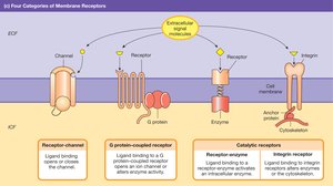
Membrane receptors are classified into four main types:
Receptor-channels: Ligand binding opens or closes the channel.
G protein-coupled receptors: Ligand binding activates a G protein, which opens an ion channel or alters enzyme activity.
Receptor-enzymes: Ligand binding activates an intracellular enzyme.
Integrin receptors: Ligand binding alters the cytoskeleton.
Signal Transduction: Second Messenger Systems
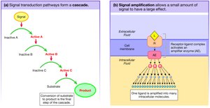
Second messenger systems amplify and distribute the signal inside the cell. The extracellular signal (first messenger) activates intracellular signaling molecules (second messengers), which can alter ion channel gating, enzyme activity, or intracellular calcium levels.
cAMP System: Ligand binds to G protein-linked receptor, activating adenylyl cyclase, which converts ATP to cAMP. cAMP activates protein kinase A, leading to protein phosphorylation.
Calcium System: Ligand activates phospholipase C, which cleaves PIP2 into DAG and IP3. IP3 increases intracellular Ca2+, which binds calmodulin and activates proteins.
Messenger System | Key Steps | Main Effect |
|---|---|---|
cAMP | Ligand → G protein → Adenylyl cyclase → cAMP → PKA | Protein phosphorylation |
Calcium | Ligand → G protein → Phospholipase C → IP3 → Ca2+ release | Protein activation via Ca2+/calmodulin |
Additional info: Second messenger cascades allow for signal amplification, where a small amount of extracellular ligand can produce a large intracellular response.
