 뒤로
뒤로Osseous Tissue Structure and Organization
스터디 가이드 - 스마트 노트
 자료에 맞춘 맞춤형 노트, 핵심 정의, 예시, 맥락을 확장해 제공합니다.
The Skeletal System: Osseous Tissue and Bone Structure
Structure of Osseous Tissue
Osseous tissue, or bone tissue, is a specialized connective tissue that forms the rigid part of the skeleton. It is composed of cells, fibers, and a mineralized matrix, providing support, protection, and facilitating movement.
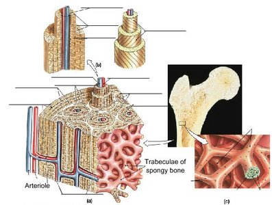
Osteons (Haversian Systems): The fundamental structural unit of compact bone, consisting of concentric rings (lamellae) surrounding a central canal.
Central Canal (Haversian Canal): The channel at the center of each osteon, containing blood vessels and nerves.
Concentric Lamellae: Layers of bony matrix arranged around the central canal.
Interstitial Lamellae: Remnants of old osteons found between newer osteons.
Circumferential Lamellae: Layers of bone matrix that encircle the entire bone, just beneath the periosteum and endosteum.
Perforating Fibers (Sharpey's Fibers): Collagen fibers that anchor the periosteum to the underlying bone.
Endosteum: A thin membrane lining the internal surfaces of bone, including the medullary cavity and trabeculae of spongy bone.
Collagen Fiber Orientation: Collagen fibers in lamellae are oriented in different directions to provide strength and resist torsion.
Example: The femur, a long bone, contains both compact bone (organized into osteons) and spongy bone (composed of trabeculae).
Vascular Supply of Bone
Bones are highly vascularized, with blood vessels running through the central canals and other channels to supply nutrients and remove waste.
Arteriole: A small artery that delivers blood to bone tissue.
Venule: A small vein that drains blood from bone tissue.
Capillary: The smallest blood vessel, facilitating exchange of nutrients and waste between blood and bone cells.
Example: Blood vessels penetrate bone through nutrient foramina and branch within the central and perforating canals.
Spongy Bone Structure
Spongy bone, also known as cancellous bone, is found at the ends of long bones and inside flat bones. It consists of a network of trabeculae, which provide structural support and house bone marrow.
Trabeculae: Thin, bony plates forming a meshwork within spongy bone.
Bone Marrow: Fills the spaces between trabeculae, involved in hematopoiesis (blood cell formation).
Example: The epiphysis of the femur contains spongy bone with trabeculae oriented along lines of stress.
Summary Table: Components of Osseous Tissue
Component | Description | Location |
|---|---|---|
Osteon | Structural unit of compact bone | Compact bone |
Central Canal | Contains blood vessels and nerves | Center of osteon |
Concentric Lamellae | Rings of matrix around central canal | Osteon |
Interstitial Lamellae | Remnants of old osteons | Between osteons |
Circumferential Lamellae | Encircle bone beneath periosteum/endosteum | Outer/inner surfaces of bone |
Perforating Fibers | Anchor periosteum to bone | Bone surface |
Endosteum | Membrane lining internal bone surfaces | Medullary cavity, trabeculae |
Arteriole | Small artery supplying bone | Central canal, periosteum |
Venule | Small vein draining bone | Central canal, periosteum |
Capillary | Smallest blood vessel | Bone tissue |
Trabeculae | Meshwork in spongy bone | Spongy bone |
Additional info: The arrangement of collagen fibers in lamellae increases bone strength and resistance to mechanical stress. The endosteum is important for bone growth, repair, and remodeling.
