 뒤로
뒤로Other Endocrine Glands and Hormone Physiology
스터디 가이드 - 스마트 노트
 자료에 맞춘 맞춤형 노트, 핵심 정의, 예시, 맥락을 확장해 제공합니다.
Other Endocrine Glands
Pineal Gland
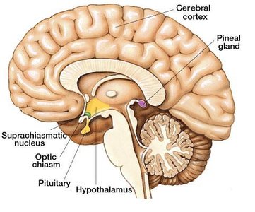
The pineal gland is a small endocrine organ located in the brain. It is most active in early childhood and its activity decreases significantly by puberty. The pineal gland produces serotonin during the day, which is converted to melatonin at night. Melatonin regulates the circadian rhythm (sleep-wake cycle) and may influence the timing of puberty in humans. Elevated melatonin levels are associated with Seasonal Affective Disorder (SAD) and Premenstrual Syndrome (PMS), and can be reduced by phototherapy. Symptoms of excess melatonin include depression, sleepiness, irritability, and carbohydrate craving.
Thymus
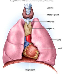
The thymus is located in the mediastinum, superior to the heart. It is most prominent in children and shrinks after puberty. The thymus secretes hormones that regulate the development and maturation of T-cells, which are essential for the immune response.
Parathyroid Glands
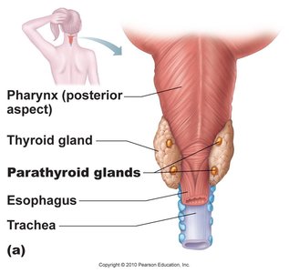
The parathyroid glands are small glands located on the posterior surface of the thyroid gland. They secrete parathyroid hormone (PTH) in response to low blood calcium levels. PTH increases blood calcium by stimulating bone resorption, increasing intestinal calcium absorption, and reducing calcium loss in urine.
Pancreas
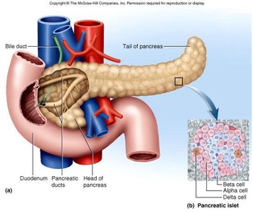
The pancreas is a retroperitoneal organ located inferior and dorsal to the stomach. It has both endocrine and exocrine functions. The endocrine portion consists of clusters called islets that produce hormones (about 2% of the organ), while the majority of the pancreas produces digestive enzymes (exocrine function).
Pancreatic Hormones
Insulin (from β cells): Secreted after meals when blood glucose levels rise. It stimulates glucose and amino acid uptake by cells and antagonizes the effects of glucagon.
Glucagon (from α cells): Secreted during fasting or low-carbohydrate diets. It stimulates the breakdown of glycogen and fat (catabolism) and antagonizes insulin.



Diabetes Mellitus
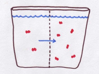
Diabetes mellitus is characterized by hyposecretion or inaction of insulin. Major symptoms include polyuria (excessive urination), polydipsia (excessive thirst), polyphagia (excessive hunger), hyperglycemia (high blood glucose), glycosuria (glucose in urine), and ketonuria (ketones in urine). Osmotic diuresis occurs as increased glucose draws water into the urine by osmosis.
Type I Diabetes Mellitus
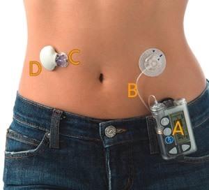
Also known as Insulin Dependent Diabetes Mellitus (IDDM), this type accounts for about 10% of diabetes cases. It is caused by autoimmune destruction of β cells, usually diagnosed around age 12. Treatment includes diet, exercise, blood glucose monitoring, and periodic insulin injections.

Type II Diabetes Mellitus
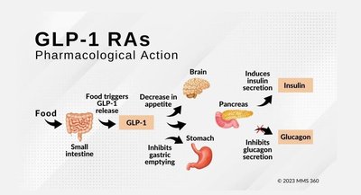
Formerly called Non-Insulin Dependent Diabetes Mellitus (NIDDM), this type represents 90% of diabetes cases. It is due to insulin resistance, where target cells fail to respond to insulin. Major risk factors include heredity, age (over 40), and obesity. Treatment involves weight loss, diet, exercise, and oral medications to improve insulin secretion or sensitivity.

Long-Term Consequences of Diabetes
Both types of diabetes can lead to vascular damage, resulting in heart and kidney disease, retinal damage (blindness), poor wound healing, and limb amputations.
Other Forms of Diabetes
Hyperinsulinism: Caused by excess insulin (injection or tumor), leading to hypoglycemia, weakness, hunger, anxiety, sweating, and increased heart rate. Severe cases can result in insulin shock (disorientation, convulsions, unconsciousness).
Gestational Diabetes: Occurs during pregnancy due to placental hormones deactivating insulin receptors. It usually resolves after birth but increases the risk of birth defects and maternal health problems, as well as future development of Type II diabetes in the mother.

Gonads
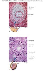
The gonads (ovaries and testes) are endocrine glands that produce sex hormones.
Ovaries: Secrete estrogens and progesterone, which regulate the development of the female reproductive system, menstrual cycle, pregnancy, and preparation of mammary glands for lactation.
Testes: Produce androgens, which regulate the development of the male reproductive system, sustain sperm production, and maintain sex drive.
Endocrine Functions of Other Organs
Heart: Releases atrial natriuretic peptide (ANP) in response to increased blood pressure, which decreases blood volume and pressure. Also produces erythropoietin (stimulates RBC production).
Stomach and Small Intestines: Secrete about 10 enteric hormones that coordinate digestive motility and secretion.
Liver: Continues synthesis of calcitriol and produces some erythropoietin.
Placenta: Secretes estrogen, progesterone, and other hormones to regulate pregnancy and stimulate fetal and mammary gland development.
Kidneys: Produce hormones involved in erythropoiesis and calcium regulation.
Hormone Physiology
Chemical Nature of Hormones
Hormones are classified based on their chemical structure:
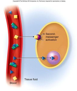
Protein-based (water-soluble) hormones: Include histamine, epinephrine, and insulin. These hormones cannot cross the plasma membrane and must bind to cell-surface receptors.
Steroid (lipid-soluble) hormones: Can penetrate the plasma membrane and bind to intracellular receptors.
Hormones circulate throughout the body but only affect target cells with specific receptors. There are two main mechanisms of hormone action: lipid-soluble and water-soluble pathways.
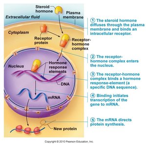
Lipid-Soluble Hormone Mode of Action
Lipid-soluble hormones (e.g., steroid hormones) diffuse through the plasma membrane and bind to intracellular receptors. The hormone-receptor complex enters the nucleus, binds to DNA, and initiates transcription of specific genes, leading to protein synthesis.
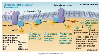
Water-Soluble Hormone Mechanism of Action
Water-soluble hormones bind to cell-surface receptors, activating second messenger systems (such as cAMP) that amplify the signal and trigger cellular responses.
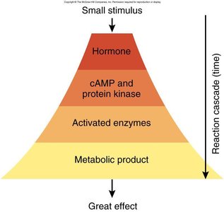
Enzyme Amplification
Hormones can trigger a cascade of enzymatic reactions, where a small stimulus leads to a large physiological effect. This process is known as enzyme amplification.
Hormone Clearance
Hormone signals must be terminated after their action. Hormones are taken up and degraded by the liver and kidneys, then excreted in bile or urine. The metabolic clearance rate (MCR) is the rate of hormone removal, and the half-life is the time required to clear 50% of the hormone from the blood.
Modulation of Target Cell Sensitivity
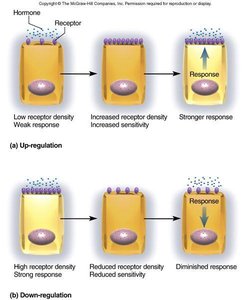
Target cells can adjust their sensitivity to hormones by changing the number of receptors:
Up-regulation: Increases receptor density, enhancing sensitivity and response.
Down-regulation: Decreases receptor density, reducing sensitivity and response.
Control of Pituitary: Feedback from Target Organs
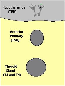
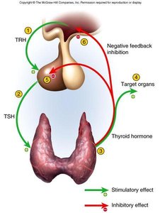
The pituitary gland is regulated by feedback from target organs. Hormones released by target organs can inhibit or stimulate further hormone release from the pituitary, maintaining homeostasis through negative feedback loops.
Stress and Adaptation
General Adaptation Syndrome
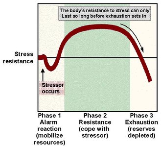
Stress is any situation that disrupts homeostasis and threatens well-being. The body's response to stress is called the General Adaptation Syndrome, which occurs in three stages:
Alarm Reaction: Mobilization of resources to face the stressor.
Resistance: Coping with the stressor and maintaining resistance.
Exhaustion: Depletion of resources, leading to decreased resistance and possible health consequences.
Paracrine Secretions
Paracrine secretions are chemical messengers that diffuse short distances to stimulate nearby cells. Unlike neurotransmitters, they are not produced by neurons, and unlike hormones, they are not transported in the blood. Examples include:
Histamine: Involved in allergic reactions and causes vasodilation.
Nitric oxide: Produced by blood vessels, causes vasodilation.
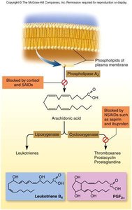
Eicosanoids: Involved in inflammation, pain, and fever; mediate allergic and inflammatory reactions, stimulate vasoconstriction and clotting, and sensitize neurons to pain.
Type | Source | Main Function |
|---|---|---|
Histamine | Mast cells | Vasodilation, allergy |
Nitric oxide | Endothelial cells | Vasodilation |
Eicosanoids | Many tissues | Inflammation, pain, fever |
Anti-inflammatory drugs can block the synthesis of eicosanoids:
Non-steroidal anti-inflammatory drugs (NSAIDs): Aspirin, Advil (block COX-1/2)
COX-2 inhibitors: Vioxx, Celebrex
Steroidal anti-inflammatory drugs: Prednisone (block phospholipase A2)
