 뒤로
뒤로Overview of Organ Systems: Structure, Function, and Laboratory Dissection
스터디 가이드 - 스마트 노트
 자료에 맞춘 맞춤형 노트, 핵심 정의, 예시, 맥락을 확장해 제공합니다.
Overview of Organ Systems
Introduction to Organ Systems
The human body is organized into multiple organ systems, each composed of related organs that work together to perform specific functions essential for survival and homeostasis. Understanding these systems is foundational for the study of anatomy and physiology, as each system contributes to the integrated function of the body as a whole.
Major Organ Systems of the Body
Each organ system has distinct structures and roles. The following table summarizes the main organ systems, their primary organs, and their major functions.
Organ System | Main Organs | Primary Functions |
|---|---|---|
Integumentary | Skin, hair, nails | Protection, temperature regulation, sensation |
Skeletal | Bones, joints | Support, movement, protection, blood cell production |
Muscular | Skeletal muscles | Movement, posture, heat production |
Nervous | Brain, spinal cord, nerves | Control, coordination, response to stimuli |
Endocrine | Glands (pituitary, thyroid, etc.) | Hormone production, regulation of metabolism and growth |
Cardiovascular | Heart, blood vessels | Transport of nutrients, gases, wastes; immune response |
Lymphatic/Immune | Lymph nodes, spleen, thymus | Fluid balance, defense against pathogens |
Respiratory | Lungs, trachea, bronchi | Gas exchange (O2/CO2) |
Digestive | Stomach, intestines, liver, pancreas | Breakdown and absorption of nutrients, waste elimination |
Urinary | Kidneys, bladder, ureters | Waste excretion, water and electrolyte balance |
Reproductive | Testes, ovaries, uterus, etc. | Production of gametes, reproduction |
Example: The cardiovascular system transports oxygen and nutrients to tissues and removes waste products, working closely with the respiratory and digestive systems.
Laboratory Dissection: Rat as a Model Organism
Purpose of Dissection
Dissection of the rat provides hands-on experience with mammalian anatomy, allowing students to observe the spatial relationships and structures of organ systems. The rat is commonly used due to its anatomical similarities to humans.
Securing and Initial Incision
Proper technique is essential for effective dissection and preservation of anatomical structures. The rat is first secured in a supine position, and an initial incision is made along the midline to expose the thoracic and abdominal cavities.
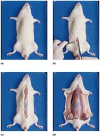
Stepwise Procedure:
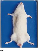
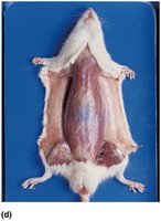
Step 1: Place the rat on its back and secure the limbs to the dissection tray.
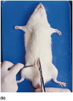
Step 2: Make an initial incision through the skin along the midline using scissors.
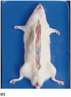
Step 3: Reflect the skin and expose the underlying musculature.
Step 4: Continue incisions to fully expose the thoracic and abdominal cavities.
Superficial Organs of the Thoracic Cavity
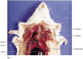
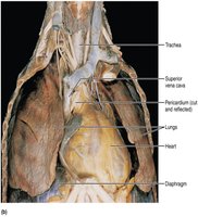
The thoracic cavity contains vital organs such as the heart and lungs, protected by the rib cage. Dissection reveals the spatial arrangement of these organs and associated structures like the trachea and thymus.
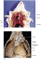
Heart: Central organ of the cardiovascular system, pumps blood throughout the body.
Lungs: Main organs of the respiratory system, responsible for gas exchange.
Trachea: Airway connecting the larynx to the bronchi.
Thymus: Lymphatic organ involved in immune function, especially in young animals.
Diaphragm: Muscular partition separating thoracic and abdominal cavities, essential for breathing.
Abdominal Organs
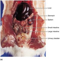
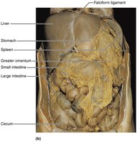
The abdominal cavity houses organs of the digestive, urinary, and reproductive systems. Dissection allows identification of the liver, stomach, intestines, spleen, and urinary bladder.
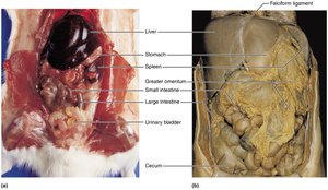
Liver: Large organ involved in metabolism, detoxification, and bile production.
Stomach: Site of mechanical and chemical digestion of food.
Spleen: Lymphatic organ involved in filtering blood and immune response.
Small Intestine: Major site of nutrient absorption.
Large Intestine: Absorbs water and forms feces.
Urinary Bladder: Stores urine prior to excretion.
Deep Structures of the Abdominopelvic Cavity
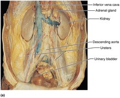
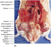
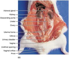
Deeper dissection reveals the kidneys, adrenal glands, ureters, and reproductive organs. These structures are essential for excretion, hormone production, and reproduction.
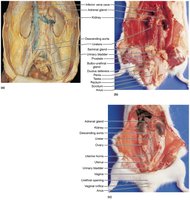
Kidneys: Filter blood to produce urine, regulate fluid and electrolyte balance.
Adrenal Glands: Produce hormones such as adrenaline and cortisol.
Ureters: Transport urine from kidneys to bladder.
Reproductive Organs: Include testes, ovaries, uterus, and associated structures.
Human Reproductive Organs
Overview of Male and Female Reproductive Anatomy
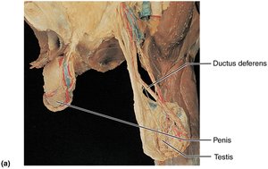
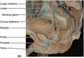
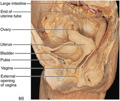
The reproductive system ensures the continuation of species through the production of gametes and support of fertilization and development. Male and female systems have distinct structures and functions.
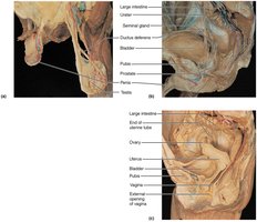
Male: Testes (sperm production), ductus deferens, prostate, penis.
Female: Ovaries (egg production), uterine tubes, uterus, vagina.
Summary Figure: Human Organ Systems
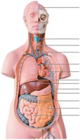
The following figure provides an overview of the major organ systems and their anatomical locations in the human body.
Additional info: Laboratory dissection is a critical skill for anatomy students, as it reinforces theoretical knowledge with practical, hands-on experience. Comparative anatomy using model organisms like the rat helps students appreciate both the similarities and differences among mammals.
