 뒤로
뒤로Plasma Membrane Structure and Membrane Potential: Study Notes for Anatomy & Physiology
스터디 가이드 - 스마트 노트
 자료에 맞춘 맞춤형 노트, 핵심 정의, 예시, 맥락을 확장해 제공합니다.
The Plasma Membrane and Membrane Potential
Body Fluid Compartments
The human body contains three main fluid compartments that are separated by membranes. These compartments are essential for maintaining homeostasis and cellular function.
Extracellular Fluid (ECF): Includes plasma (the fluid portion of blood) and interstitial fluid (fluid surrounding most cells).
Intracellular Fluid (ICF): The fluid within cells, which constitutes the majority of body water.
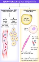
Compartment Separation by Membranes
Fluid compartments are separated by biological membranes, which serve as barriers and regulate the movement of substances between compartments.
Tissue membranes: Composed of layers of connective tissue and cells.
Cell membranes: Phospholipid bilayers that create cell boundaries.
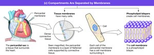
Biological Membranes: Structure and Function
Fluid Mosaic Model
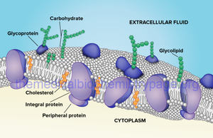
The plasma membrane is described by the fluid mosaic model, which highlights its dynamic nature and diverse components. The membrane separates the cell from its environment, encloses intracellular contents, and acts as a mechanical barrier.
Lipid bilayer: Provides the basic structure and fluidity.
Proteins: Embedded or attached, serving various functions.
Carbohydrates: Attached to proteins and lipids, important for cell recognition.
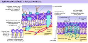
Lipid Bilayer
The lipid bilayer forms the fundamental structure of the plasma membrane. It consists mainly of phospholipids, which are amphipathic molecules with hydrophilic heads and hydrophobic tails. Cholesterol is interspersed within the bilayer, contributing to membrane fluidity and stability.
Hydrophobic interior: Acts as a barrier to water-soluble substances.
Fluidity: Maintained by cholesterol, preventing phospholipid tails from packing tightly.
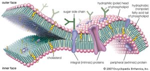
Membrane Proteins
Proteins in the plasma membrane are classified as integral (spanning the membrane) or peripheral (attached to the surface). They perform critical functions such as transport, signaling, and cell adhesion.
Channels: Allow passage of ions and water.
Carrier molecules: Facilitate transport of larger or polar molecules.
Receptors: Bind signaling molecules.
Enzymes: Catalyze reactions.
Cell adhesion molecules (CAMs): Link cells together.
Recognition: Identify 'self' cells.
Membrane Carbohydrates
Carbohydrates are found on the outer surface of the plasma membrane, attached to proteins (glycoproteins) or lipids (glycolipids). They play a key role in cell recognition and immune response.
Cell recognition: Helps distinguish self from non-self.
Cell-to-Cell Adhesion and Junctions
Extracellular Matrix and Cell Adhesion Molecules
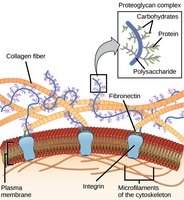
Cells are bound together into tissues and organs by the extracellular matrix and cell adhesion molecules (CAMs). The matrix is a meshwork of fibrous proteins and polysaccharides, facilitating diffusion and structural support.
CAMs: Membrane proteins that link cells via hooks and loops.
Specialized Cell Junctions
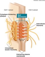
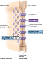
Specialized junctions provide structural integrity and communication between cells.
Desmosomes: Adhering junctions found in tissues subject to stretching.
Tight junctions: Seal off passageways between adjacent cells.
Gap junctions: Connect cytoplasm of adjacent cells, allowing communication.
Membrane Transport
Selective Permeability
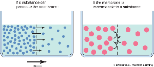
The plasma membrane is selectively permeable, allowing certain substances to pass while restricting others. Permeability depends on solubility, size, and method of transport.
Nonpolar and uncharged: Can cross easily.
Polar and charged: Require assistance.
Small molecules: More likely to pass.
Large molecules: Usually blocked.
Unassisted Membrane Transport
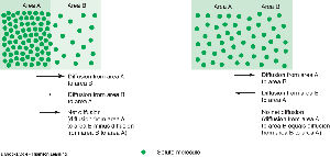
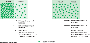
Some molecules can cross the membrane without assistance, primarily through diffusion down a concentration gradient.
Diffusion: Movement from high to low concentration.
Equilibrium: Achieved when net movement ceases.
Fick's Law of Diffusion
The rate of diffusion across a membrane is described by Fick's Law:
Increase rate: Greater concentration gradient (ΔC), higher permeability (P), larger surface area (A).
Decrease rate: Higher molecular weight (MW), greater distance (ΔX).
Equation:
Where J is the rate of diffusion, P is permeability, A is surface area, and ΔC is the concentration gradient.
Assisted Membrane Transport
Large, polar, or charged molecules require assistance to cross the plasma membrane. Mechanisms include facilitated diffusion, osmosis, active transport, and vesicular transport.
Channel proteins: Form open passageways for ions and water.
Carrier proteins: Change conformation to move molecules.
Channel Proteins
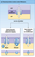
Channel proteins create water-filled pores in the membrane, allowing specific ions or water to pass. Channels can be open (leaky) or gated (responding to signals).
Types: Chemically gated, voltage-gated, mechanically gated.
Carrier Proteins
Carrier proteins bind to solutes and change shape to transport them across the membrane. They can move substances via uniport (single molecule), symport (co-transport in same direction), or antiport (exchange in opposite directions).
Facilitated diffusion: Down concentration gradient, no energy required.
Active transport: Against concentration gradient, requires energy.
Osmosis and Osmolarity
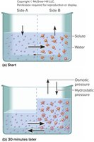
Osmosis is the movement of water across a membrane, driven by solute concentration differences. Membranes are impermeable to water without aquaporins. Osmolarity refers to the concentration of solutes, influencing water movement and osmotic pressure.
Water moves: Toward higher solute concentration.
Osmotic pressure: Generated by water movement.
Facilitated Diffusion vs. Active Transport
Both processes require helper proteins, but differ in energy requirements and direction of transport.
Facilitated diffusion: Down concentration or electrical gradient; uses channels and carriers.
Active transport: Against gradient; requires energy; uses carrier proteins only (e.g., Na+/K+ pump).
Vesicular Transport
Vesicular transport involves moving material into or out of the cell wrapped in membrane vesicles. Types include endocytosis (pinocytosis, phagocytosis) and exocytosis.
Passive Movement Down an Electrical Gradient
Charged particles move according to electrochemical gradients, which combine concentration and charge differences across the membrane.
Membrane Potential
The Resting Membrane Potential
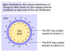
Cells maintain a resting membrane potential due to unequal distribution of ions. The intracellular fluid (ICF) is negatively charged relative to the extracellular fluid (ECF), which is positively charged.
Membrane Potential: Charge Attraction and Measurement
Membrane potential is the voltage difference across the plasma membrane, measured in millivolts (mV). It represents potential energy due to separated charges.
Membrane Potentials of Cells
Every cell has a resting membrane potential (Vrest), determined by ion distribution and permeability. The Na+/K+ pump and leaky channels maintain this potential.
Na+: Higher outside the cell.
K+: Higher inside the cell.
A-: Anions remain inside the cell.
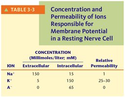
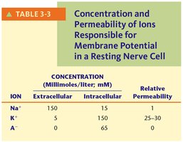
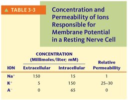
Ion | Extracellular (mM) | Intracellular (mM) | Relative Permeability |
|---|---|---|---|
Na+ | 150 | 15 | 1 |
K+ | 5 | 150 | 25–30 |
A- | 0 | 65 | 0 |
Equilibrium Potential and Nernst Equation
The equilibrium potential for an ion is the voltage at which there is no net movement of that ion across the membrane. It is calculated using the Nernst equation:
Example for K+:
Example for Na+:
Goldman Equation
The Goldman equation calculates the steady-state membrane potential considering multiple ions and their permeabilities:
PK: Permeability of K+ (set to 1)
PNa: Permeability of Na+ (set to 0.04)
Net Force and Ion Movement
The net force on an ion is determined by the difference between membrane potential (Vm) and equilibrium potential (Ex):
If Vm - Ex is positive, a positive ion moves out of the cell.
If Vm - Ex is negative, a positive ion moves into the cell.
If Vm - Ex is zero, there is no net force.
Specialized Use of Membrane Potential
Nerve and muscle cells can rapidly alter their membrane potential in response to stimulation, producing nerve impulses and triggering muscle contraction.
Example: Action potentials in neurons and muscle cells are generated by transient changes in ion permeability.
