뒤로
뒤로Structure and Accessory Structures of the Eye
스터디 가이드 - 스마트 노트
 자료에 맞춘 맞춤형 노트, 핵심 정의, 예시, 맥락을 확장해 제공합니다.
Accessory Structures of the Eye
Lacrimal Apparatus
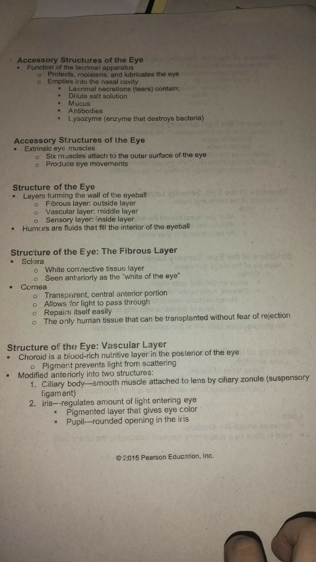
The lacrimal apparatus is responsible for producing and draining tears, which serve to protect, moisten, and lubricate the eye. Tears are secreted by the lacrimal glands and emptied into the nasal cavity.
Functions: Protects, moistens, and lubricates the eye
Lacrimal secretions (tears) contain:
Dilute salt solution
Mucus
Antibodies
Lysozyme (an enzyme that destroys bacteria)
Extrinsic Eye Muscles
Six extrinsic eye muscles attach to the outer surface of the eye, allowing for a wide range of eye movements. These muscles are essential for coordinated and precise movement of the eyeball.
Six muscles attach to the outer surface of the eye
Produce eye movements
Structure of the Eye
Layers of the Eyeball
The wall of the eyeball is composed of three main layers, each with distinct functions and structures. Additionally, the interior of the eye is filled with fluids known as humors.
Fibrous layer: Outside layer
Vascular layer: Middle layer
Sensory layer: Inside layer
Humors: Fluids that fill the interior of the eyeball
The Fibrous Layer
The fibrous layer is the outermost layer of the eye, providing structure and protection. It consists of the sclera and the cornea.
Sclera:
White connective tissue layer
Seen anteriorly as the "white of the eye"
Cornea:
Transparent, central anterior portion
Allows for light to pass through
Repairs itself easily
The only human tissue that can be transplanted without fear of rejection
The Vascular Layer
The vascular layer is the middle layer of the eye, rich in blood vessels and pigment. It nourishes the eye and prevents light from scattering within the eyeball.
Choroid: Blood-rich nutritive layer in the posterior of the eye
Pigment prevents light from scattering
Modified anteriorly into two structures:
Ciliary body: Smooth muscle attached to the lens by the ciliary zonule (suspensory ligament)
Iris: Regulates the amount of light entering the eye
Pigmented layer that gives eye color
Pupil: Rounded opening in the iris