 뒤로
뒤로Structure of the Eye: Sensory Layer and Lens
스터디 가이드 - 스마트 노트
 자료에 맞춘 맞춤형 노트, 핵심 정의, 예시, 맥락을 확장해 제공합니다.
Structure of the Eye: Sensory Layer
Overview of the Retina
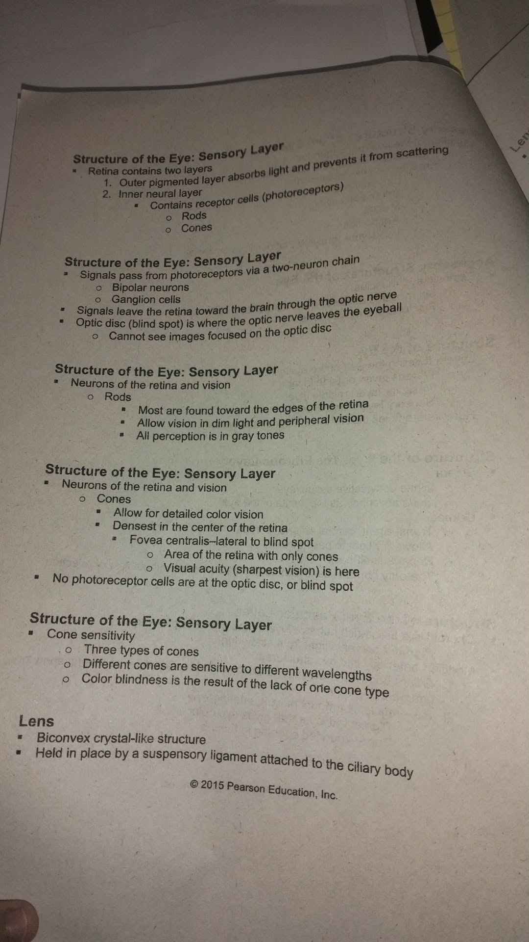
The retina is the sensory layer of the eye responsible for detecting light and initiating the process of vision. It is composed of two main layers that work together to absorb light and transmit visual information to the brain.
Outer pigmented layer: Absorbs light and prevents it from scattering within the eye, which helps to produce a clear image.
Inner neural layer: Contains receptor cells known as photoreceptors, which are specialized neurons that respond to light. There are two main types of photoreceptors:
Rods
Cones
Neural Pathways in the Retina
Signals from photoreceptors are transmitted through a two-neuron chain to the brain:
Bipolar neurons
Ganglion cells
These signals leave the retina via the optic nerve. The optic disc (also known as the blind spot) is the region where the optic nerve exits the eyeball. No photoreceptor cells are present at the optic disc, so images focused here cannot be seen.
Photoreceptors: Rods and Cones
Rods:
Located primarily at the edges of the retina.
Enable vision in dim light and are responsible for peripheral vision.
All perception by rods is in shades of gray (no color detection).
Cones:
Allow for detailed color vision.
Most densely packed in the center of the retina, especially in the fovea centralis (located lateral to the blind spot).
The fovea centralis contains only cones and is the region of the retina with the highest visual acuity (sharpest vision).
No photoreceptor cells are present at the optic disc (blind spot).
Cone Sensitivity and Color Vision
Cones are responsible for color vision and visual acuity. There are three types of cones, each sensitive to different wavelengths of light (corresponding to red, green, and blue light).
Three types of cones: Each type is maximally sensitive to a specific range of wavelengths.
Color blindness: Results from the absence or malfunction of one or more types of cones, leading to difficulty distinguishing certain colors.
Lens
Structure and Function
The lens is a transparent, biconvex, crystal-like structure that focuses light onto the retina. It is held in place by a suspensory ligament attached to the ciliary body, allowing it to change shape for focusing (accommodation).
Biconvex shape: Allows the lens to bend light rays to focus them on the retina.
Suspensory ligament: Connects the lens to the ciliary body, enabling adjustments in lens shape for near and distant vision.
Additional info: The fovea centralis is the region of the retina with the highest density of cones and is critical for tasks requiring detailed vision, such as reading and recognizing faces. The process by which the lens changes shape to focus on objects at different distances is called accommodation.
