 뒤로
뒤로Study Guide: The Cardiovascular System – Heart and Blood Vessels
스터디 가이드 - 스마트 노트
 자료에 맞춘 맞춤형 노트, 핵심 정의, 예시, 맥락을 확장해 제공합니다.
Cardiovascular System Overview
Introduction
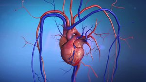
The cardiovascular system is responsible for the transport of blood, nutrients, gases, and wastes throughout the body. It consists of the heart, blood vessels, and blood. Understanding the structure and function of these components is essential for comprehending human physiology.
Blood Vessels
Types of Blood Vessels
Blood vessels are classified into three main types based on their function and structure:
Arteries: Carry blood away from the heart to tissues. Usually transport oxygenated blood, except in pulmonary and fetal circulation. Arteries have thicker walls due to a prominent muscle layer.
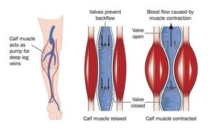
Veins: Carry blood toward the heart from tissues. Usually transport deoxygenated blood, except in pulmonary and fetal circulation. Veins contain valves to prevent backflow.
Capillaries: Microscopic vessels that connect arterioles and venules. They facilitate gaseous exchange and consist of a single layer of epithelial cells.
Structure of Blood Vessel Walls (Tunics)
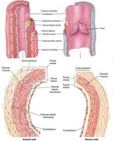
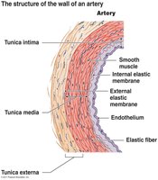
Blood vessel walls are composed of three layers, known as tunics:
Tunica externa: The outer layer, made of elastic and collagen fibers.
Tunica media: The middle layer, consisting of smooth muscle and elastic tissue.
Tunica intima: The inner layer, composed of endothelium.
Venous tunics are named the same but are thinner than arteries, contain less muscle, lack elastic fibers, and have valves.
Heart Structure
Layers of the Heart Wall
The heart wall consists of three distinct layers:
Epicardium: The outer layer, also known as the visceral pericardium.
Myocardium: The middle layer, composed of cardiac muscle (myocytes).
Endocardium: The inner layer, made of endothelium that lines the chambers and vessels.
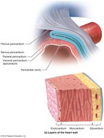
Histology of the Heart
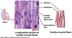
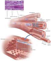
Cardiac muscle tissue forms the bulk of the heart wall. It is characterized by:
Striated, involuntary, branched, and cylindrical cells.
Presence of intercalated discs, which join adjacent cardiac muscle cells.
Intercalated discs contain desmosomes (hold cells together) and gap junctions (allow rapid ion passage and communication).
Cardiac muscle cells contain myoglobin (for O2 storage) and abundant mitochondria (for ATP production).
Heart Chambers
Overview of Chambers
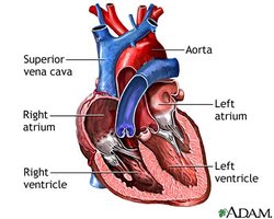
The heart has four chambers:
Atria: Two upper chambers (right and left atrium).
Ventricles: Two lower chambers (right and left ventricle).
Right Atrium
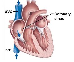
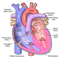
The right atrium receives deoxygenated blood from three sources:
Superior vena cava (SVC)
Inferior vena cava (IVC)
Coronary sinus
Blood passes from the right atrium to the right ventricle through the tricuspid valve.
Right Ventricle
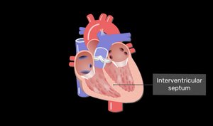
The right ventricle contains trabeculae carneae, which are raised bundles of cardiac fibers involved in the conduction system. The right and left ventricles are separated by the interventricular septum.
Blood Flow from Right Ventricle
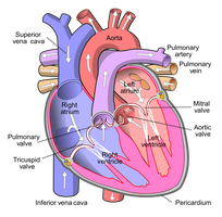
Blood passes from the right ventricle through the pulmonary semilunar valve into the pulmonary trunk, which divides into right and left pulmonary arteries leading to the lungs.
Left Atrium
The left atrium receives oxygenated blood from the lungs via the pulmonary veins. Blood passes from the left atrium to the left ventricle through the bicuspid (mitral) valve.
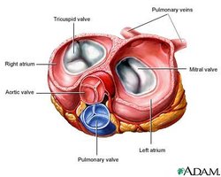
Left Ventricle
Oxygenated blood passes from the left ventricle through the aortic semilunar valve into the ascending aorta, which distributes blood to the body tissues. The left ventricle also contains trabeculae carneae.
Heart Valves
Chordae Tendineae and Papillary Muscles
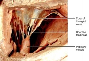
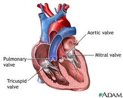
Chordae tendineae are fibrous cords that connect atrioventricular (AV) valves to papillary muscles. Papillary muscles contract to close AV valves and prevent backflow of blood into the atria.
Types of Heart Valves
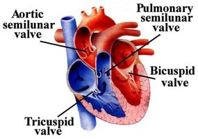
Semilunar Valves: Aortic and pulmonary valves. Blood flows into the aorta and pulmonary trunk as ventricles contract and valves open.
Atrioventricular Valves: Bicuspid (mitral) and tricuspid valves. Blood flows from atria into ventricles through AV valves; as ventricles contract, valve cusps close to prevent backflow.
Coronary Circulation
Coronary Arteries
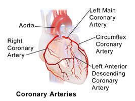
The myocardium is too thick for oxygen and nutrients to diffuse from the chambers, so the heart is supplied by coronary circulation:
Right coronary artery: Supplies right atrium and ventricle.
Left coronary artery: Branches into the anterior interventricular artery (LAD) and circumflex artery, supplying the left atrium and ventricle.
Coronary Veins
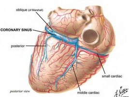
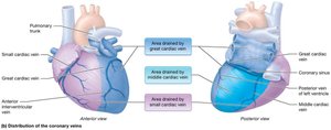
Most heart veins empty into the coronary sinus, which drains into the posterior right atrium. Major veins include:
Great cardiac vein
Small cardiac vein
Middle cardiac vein
Coronary Artery Disease (CAD) and Myocardial Infarction (MI)
CAD is caused by the build-up of fatty plaques in coronary arteries, leading to decreased blood flow and myocardial ischemia. Symptoms include chest pain (angina pectoris), shortness of breath, and other signs. MI (heart attack) occurs when a plaque ruptures and a clot forms, obstructing blood flow and causing tissue death.
LAD (left anterior descending artery) is the most common site of blockage, known as the "widow maker".
Symptoms may differ in women, often presenting as back, jaw, or arm pain.
Blood Flow Through the Heart
Pathway of Blood
Once oxygen is distributed to tissues, red blood cells pick up carbon dioxide and return it to the right atrium via the SVC, IVC, and coronary sinus. Blood then follows a specific pathway through the heart chambers and valves.
Internal Sheep Heart Anatomy
Comparative Anatomy
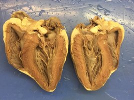
Sheep heart anatomy is often used in laboratory settings to study internal heart structures, as it closely resembles human heart anatomy.
Summary Table: Blood Vessel and Heart Wall Layers
Structure | Layers | Main Features |
|---|---|---|
Artery | Tunica externa, Tunica media, Tunica intima | Thick muscle layer, elastic fibers |
Vein | Tunica externa, Tunica media, Tunica intima | Thinner wall, valves, less muscle |
Heart Wall | Epicardium, Myocardium, Endocardium | Outer (visceral pericardium), middle (cardiac muscle), inner (endothelium) |
Key Equations
Blood Flow Equation
Blood flow through vessels is governed by the following equation:
Q: Blood flow
ΔP: Pressure difference
R: Resistance
Cardiac Output Equation
Cardiac output is the volume of blood pumped by the heart per minute:
CO: Cardiac output
HR: Heart rate
SV: Stroke volume
Additional info:
Cardiac muscle cells are highly specialized for continuous rhythmic contraction.
Coronary circulation is essential for maintaining heart function and preventing ischemic damage.
Blood vessel structure is closely related to function, with arteries designed for high-pressure transport and veins for low-pressure return.
