 뒤로
뒤로Study Guide: The Circulatory System – Blood
스터디 가이드 - 스마트 노트
 자료에 맞춘 맞춤형 노트, 핵심 정의, 예시, 맥락을 확장해 제공합니다.
The Circulatory System: Blood
Introduction
The circulatory system is essential for transporting substances throughout the body, maintaining homeostasis, and defending against disease. Blood, as a key component, serves multiple functions and is composed of plasma and formed elements.
Functions of the Circulatory System
The circulatory system consists of the heart, blood vessels, and blood. Its primary functions include:
Transport: Delivers oxygen, nutrients, hormones, and removes waste products.
Protection: Defends against pathogens and prevents blood loss through clotting.
Regulation: Maintains pH, temperature, and fluid balance.
Components and General Properties of Blood
Blood is a liquid connective tissue composed of plasma and formed elements. It has unique physical and chemical properties:
Volume: 4-6 liters in adults
Body weight: ~8%
Temperature: 38°C
Viscosity: 5 times more viscous than water
Prevents blood from leaking; if too thin, it will not clot, bp will lower
if too thick, dehydrated, heart works harder sto pump blood
pH: 7.35 - 7.45 (acidosis < 7.35, alkalosis > 7.45)
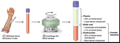
Blood Composition
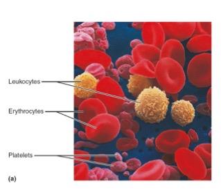
Blood consists of approximately 55% plasma and 45% formed elements. The formed elements include erythrocytes (red blood cells), leukocytes (white blood cells), and platelets.
Plasma: The liquid portion, mostly water, proteins, and solutes.
Formed Elements: Cellular components including RBCs, WBCs, and platelets.
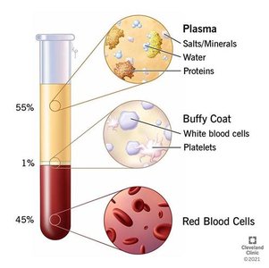
Blood Plasma
Plasma is a mixture of water (92%), proteins (7%), and other solutes (1%). Plasma proteins are the most abundant solute and play critical roles in clotting, immune defense, and transport.
Albumin: Smallest and most abundant; maintains viscosity and osmolarity.
contributes to thickness and allows blood to pass through
Globulins: Involved in immune defense and transport
Fibrinogen: Essential for blood clotting.
Liver supplies most proteins
Production of Blood (Hematopoiesis)
Blood cells are continuously produced through hematopoiesis. Daily production includes billions of platelets, RBCs, and WBCs. All formed elements originate from hematopoietic stem cells.
Erythropoiesis: Production of red blood cells.
Leukopoiesis: Production of white blood cells.
Thrombopoiesis: Production of platelets.
Erythrocytes (Red Blood Cells)
Quantity and Structure
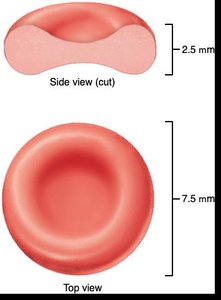
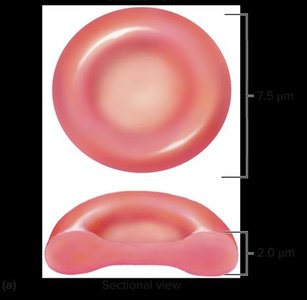
Erythrocytes are the most abundant formed element and are critical for oxygen transport. Their bi-concave disc shape increases surface area for gas exchange. They lack a nucleus and most organelles, which allows more room for hemoglobin but prevents self-repair.
Hematocrit: Clinical measurement of RBC percentage in blood.
Shape: Bi-concave disc, 7.5 μm diameter, 2.0-2.5 μm thick, no nucleus
Hemoglobin
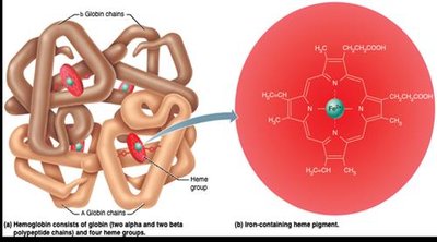
Hemoglobin is a red, iron-containing protein responsible for oxygen transport. Each iron atom binds one oxygen molecule. RBCs lack mitochondria and rely on anaerobic fermentation for ATP production.
Structure: Four globin chains, each with a heme group.
Function: Oxygen and carbon dioxide transport.
Life Cycle of Erythrocytes
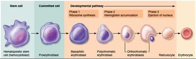
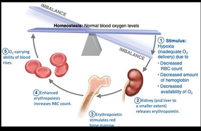
RBCs live for about 120 days. Erythropoiesis takes 3-4 days, starting from hematopoietic stem cells. Old RBCs rupture (hemolysis) and their components are recycled.
Birth: Erythropoiesis in bone marrow.
Death: Hemolysis in capillaries.
Sickle-Cell Anemia
Sickle-cell anemia is caused by a genetic mutation in hemoglobin, resulting in sickle-shaped RBCs. This affects oxygen transport and can cause blockages in blood vessels.
Genetics: Two copies of the HbS gene cause the disease.
Treatments: Focus on symptom management and prevention of complications.
Leukocytes (White Blood Cells)
Structure and Function
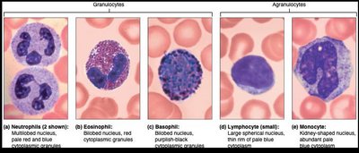
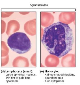
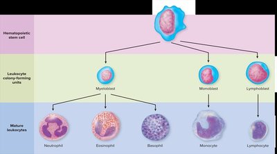
Leukocytes are the body's defense cells, residing mainly in connective tissues but traveling via the bloodstream. They are classified as granulocytes or agranulocytes based on the presence of granules.
Granulocytes: Neutrophils, eosinophils, basophils.
Agranulocytes: Monocytes, lymphocytes.
Leukocyte Life Cycle
Leukopoiesis is the process of WBC production, starting from hematopoietic stem cells. Leukocytes have varied lifespans and functions depending on their type.
Leukocyte Disorders
Disorders include leukemias (overproduction of abnormal WBCs) and infectious mononucleosis (viral disease causing excess lymphocytes).
Leukemia: Cancerous proliferation of abnormal WBCs.
Infectious mononucleosis: Caused by Epstein-Barr virus, results in excess lymphocytes.
Platelets and Control of Bleeding
Platelet Structure and Function
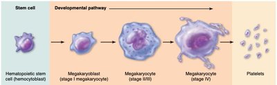
Platelets are small fragments of megakaryocytes and play a crucial role in blood clotting. They are the second most abundant formed element after RBCs.
Origin: Megakaryocytes in bone marrow.
Function: Initiate clotting and repair damaged vessels.
Control of Bleeding (Hemostasis)
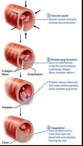
Hemostasis is the process of stopping bleeding, involving three stages: vascular spasm, platelet plug formation, and coagulation.
Vascular spasm: Smooth muscle contracts to reduce blood flow.
Platelet plug: Platelets adhere to exposed collagen fibers.
Coagulation: Fibrin forms a mesh that traps blood cells.
Agglutination vs. Coagulation
Both involve cells sticking together, but mechanisms differ:
Coagulation: Formed elements stuck by fibrin.
Agglutination: RBCs stuck by antibodies.
Blood Types and Transfusion
Blood Typing
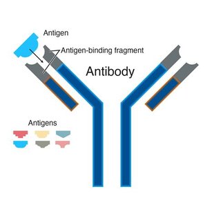
Blood types are determined by antigens (cell surface markers) and antibodies. Agglutination occurs when antibodies bind to antigens on RBCs, causing them to stick together.
Antigens: Identify self vs. foreign cells.
Antibodies: Recognize and bind to antigens.
ABO Blood Types
The ABO system classifies blood based on the presence of A and B antigens:
Type | Antigens | Antibodies |
|---|---|---|
A | A | B |
B | B | A |
AB | A and B | None |
O | None | A and B |
Universal Donor and Recipient
Type O is the universal donor (no antigens), and type AB is the universal recipient (no antibodies). Only RBCs are transfused, not plasma.
Rh Blood Typing
The Rh system is based on the presence or absence of the Rh antigen. Blood types are denoted as positive or negative (e.g., A+, A-). Rh incompatibility can cause problems in pregnancy.
Rh positive: Rh antigen present.
Rh negative: Rh antigen absent; antibodies produced only if exposed.
Summary Table: Blood Components
Component | Percentage | Main Function |
|---|---|---|
Plasma | ~55% | Transport, regulation |
Red Blood Cells (Erythrocytes) | ~45% | Oxygen transport |
White Blood Cells (Leukocytes) | <1% | Immune defense |
Platelets | <1% | Clotting |
Additional info: Academic context was added to clarify the functions, structure, and clinical relevance of blood components, as well as the mechanisms of hemostasis and blood typing.
