 뒤로
뒤로Study Guide: The Reproductive System (Anatomy & Physiology)
스터디 가이드 - 스마트 노트
 자료에 맞춘 맞춤형 노트, 핵심 정의, 예시, 맥락을 확장해 제공합니다.
The Reproductive System
Overview
The reproductive system is responsible for producing, storing, and transporting gametes, as well as secreting hormones that regulate reproductive functions and secondary sex characteristics. It consists of primary sex organs (gonads) and accessory structures.
Primary sex organs: Testes (male), Ovaries (female)
Accessory organs: Ducts, glands, and external genitalia
Sex hormones: Androgens (male), Estrogens and progesterone (female)
Anatomy of the Male Reproductive System
Male Reproductive Structures
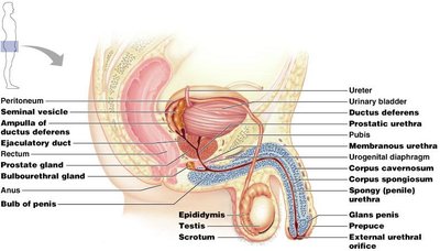
The male reproductive system includes the testes, ducts, accessory glands, and external genitalia. The testes produce sperm and hormones, while ducts and glands facilitate sperm transport and maturation.
Testes: Located within the scrotum; site of sperm production
Duct system: Epididymis → Ductus deferens → Ejaculatory duct → Urethra
Accessory glands: Seminal glands, prostate, bulbo-urethral glands
External genitalia: Penis and scrotum
The Scrotum
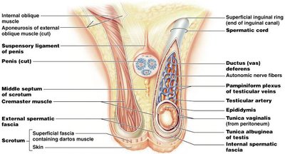
The scrotum is a sac of skin and superficial fascia that houses the testes. It maintains a temperature lower than the core body temperature, which is essential for sperm production.
Dartos muscle: Smooth muscle that wrinkles scrotal skin and pulls scrotum closer to the body
Cremaster muscle: Skeletal muscle bands that elevate the testes
The Testes
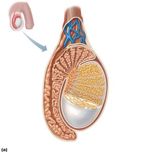
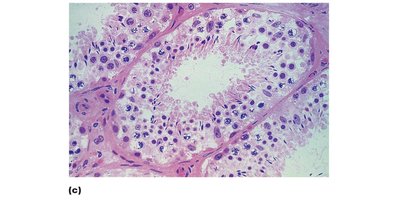
Each testis is surrounded by two tunics: the tunica vaginalis (outer) and tunica albuginea (inner). The testis is divided into lobules containing seminiferous tubules, the site of sperm production.
Tunica vaginalis: Derived from peritoneum
Tunica albuginea: Fibrous capsule
Seminiferous tubules: Site of spermatogenesis
Interstitial endocrine cells: Produce testosterone
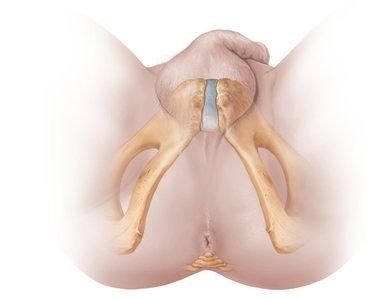
Male Perineum
The perineum is a diamond-shaped region that suspends the scrotum and contains the root of the penis and anus.
Penis: Structure and Function
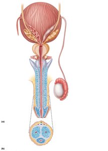
The penis is the male copulatory organ, designed to deliver sperm into the female reproductive tract. It consists of the root, shaft, glans penis, and prepuce (foreskin).
Corpora cavernosa: Paired dorsal erectile bodies
Corpus spongiosum: Surrounds urethra and forms glans and bulb
Erection: Erectile tissue fills with blood, causing rigidity
Cell Growth and Reproduction
The Cell Cycle
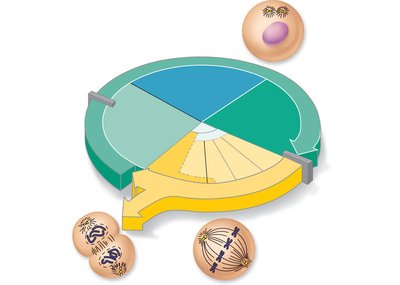
The cell cycle describes the series of events from cell formation to cell division. It includes interphase (G1, S, G2) and the mitotic phase (mitosis and cytokinesis).
Interphase: Cell growth and DNA replication
Mitosis: Nuclear division (prophase, metaphase, anaphase, telophase)
Cytokinesis: Division of cytoplasm
DNA Replication
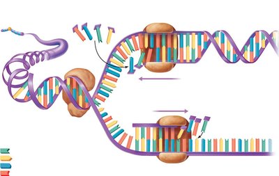
DNA replication occurs during the S phase of interphase. Helicase unwinds the DNA, and DNA polymerase synthesizes new strands using the original as a template.
Leading strand: Synthesized continuously
Lagging strand: Synthesized in segments, joined by DNA ligase
Mitosis: Stages
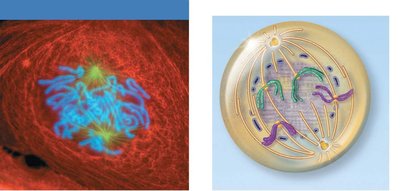
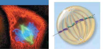
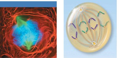
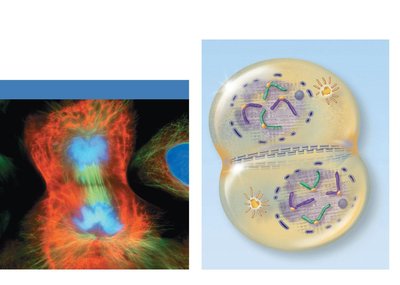
Mitosis is the process of nuclear division, essential for growth and tissue repair. It consists of four stages:
Prophase: Chromosomes condense, spindle forms
Metaphase: Chromosomes align at the metaphase plate
Anaphase: Sister chromatids separate
Telophase: Nuclear envelope reforms, chromosomes decondense
Spermatogenesis
Overview of Spermatogenesis
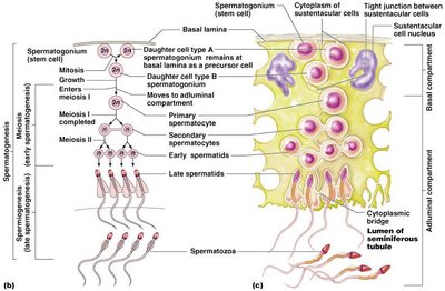
Spermatogenesis is the process of sperm production in the seminiferous tubules. It involves mitosis, meiosis, and spermiogenesis.
Diploid (2n): 46 chromosomes
Haploid (n): 23 chromosomes (gametes)
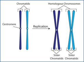
Stages of Spermatogenesis
Spermatogenic cells undergo a series of divisions and transformations:
Mitosis: Spermatogonia divide to form primary spermatocytes
Meiosis I: Primary spermatocyte (2n) → two secondary spermatocytes (n)
Meiosis II: Secondary spermatocytes (n) → spermatids (n)
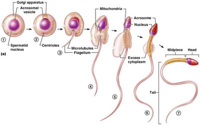
Spermiogenesis: Spermatids mature into spermatozoa
Role of Sustentocytes
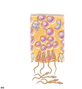
Sustentocytes (Sertoli cells) support and nourish developing sperm cells. Their tight junctions form the blood-testis barrier, protecting sperm from immune attack.
Basal compartment: Spermatogonia and primary spermatocytes
Adluminal compartment: Meiotically active cells and tubule lumen
Hormonal Regulation of Male Reproductive Function
Hypothalamic-Pituitary-Gonadal (HPG) Axis
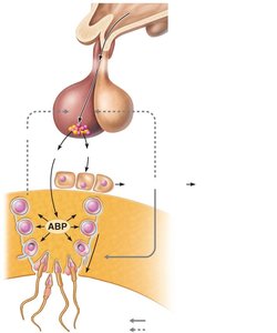
The HPG axis regulates testicular function through a sequence of hormonal interactions:
GnRH: Stimulates release of FSH and LH from anterior pituitary
FSH: Stimulates sustentocytes to release androgen-binding protein (ABP)
LH: Stimulates interstitial cells to produce testosterone
Testosterone and inhibin: Provide negative feedback to hypothalamus and pituitary
Testosterone Activity
Testosterone is synthesized from cholesterol and must be converted to dihydrotestosterone (DHT) or estrogen in certain tissues to exert its effects. It is essential for the development and maintenance of male secondary sex characteristics.
Effects: Growth of facial/body hair, deepening of voice, increased muscle mass, libido
Anatomy of the Female Reproductive System
Female Reproductive Structures
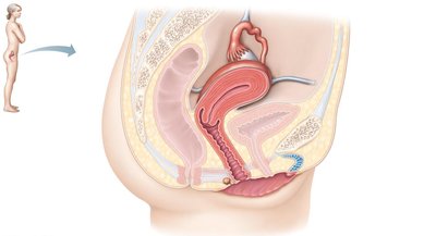
The female reproductive system includes the ovaries, uterine tubes, uterus, vagina, and external genitalia. The ovaries produce ova and hormones.
Ovaries: Primary female reproductive organs
Accessory ducts: Uterine tubes, uterus, vagina
External genitalia: Vulva (mons pubis, labia majora/minora, clitoris)
The Ovaries
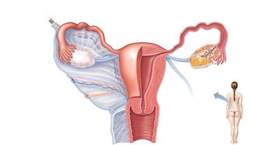
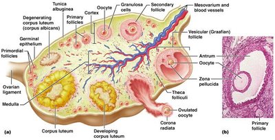
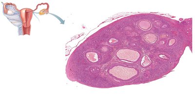
Ovaries are held in place by ligaments and are surrounded by a fibrous tunica albuginea. The cortex contains ovarian follicles at various stages of development.
Ovarian follicles: Oocyte surrounded by follicle or granulosa cells
Stages: Primordial, primary, secondary, vesicular (antral/tertiary) follicle
Ovulation: Ejection of oocyte from mature follicle
Corpus luteum: Ruptured follicle after ovulation
Female Duct System
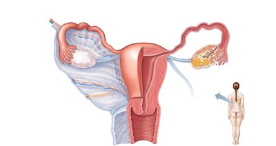
The female duct system receives the ovulated oocyte and provides a site for fertilization and development.
Uterine tubes: Carry oocyte to uterus; fimbriae create currents to move oocyte
Uterus: Muscular organ for embryo development
Vagina: Passageway for birth, menstrual flow, and copulation
Uterine Wall and Vascular Supply
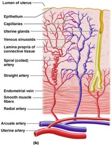
The uterine wall consists of three layers: perimetrium, myometrium, and endometrium. The endometrium undergoes cyclic changes and is supplied by a complex vascular network.
Perimetrium: Outermost serous layer
Myometrium: Smooth muscle layer
Endometrium: Mucosal lining; stratum functionalis and stratum basalis
Vagina and External Genitalia
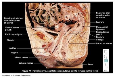
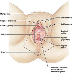
The vagina is a muscular tube that provides a passageway for birth and menstrual flow. The external genitalia (vulva) include the mons pubis, labia majora/minora, clitoris, and vestibule.
Hymen: Incomplete partition near vaginal orifice
Clitoris: Erectile tissue homologous to the penis
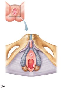
Perineum: Diamond-shaped region between pubic arch and coccyx
Mammary Glands
Mammary glands are modified sweat glands that produce milk in lactating women. They consist of lobes, lobules, ducts, and sinuses.
Areola: Pigmented skin around nipple
Suspensory ligaments: Attach breast to muscle
Lactiferous ducts: Transport milk to nipple
Oogenesis and the Ovarian Cycle
Oogenesis
Oogenesis is the production of female gametes (ova). It begins in the fetal period and continues until menopause.
Oogonia: Stem cells multiply by mitosis
Primary oocytes: Begin meiosis, stall in prophase I
Secondary oocyte: Completes meiosis I each month; arrests in metaphase II
Ovum: Formed only if fertilization occurs
Ovarian Cycle
The ovarian cycle is a monthly series of events associated with the maturation of an egg. It consists of the follicular phase, ovulation, and luteal phase.
Follicular phase: Follicle growth (days 1–14)
Ovulation: Release of secondary oocyte
Luteal phase: Corpus luteum activity (days 14–28)
Hormonal Regulation of the Ovarian Cycle
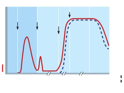
Hormone interactions produce cyclic events in the ovaries. GnRH stimulates FSH and LH release, which promote follicle growth and hormone production. Estrogen and progesterone regulate the cycle via feedback mechanisms.
Uterine (Menstrual) Cycle
Phases of the Uterine Cycle
The uterine cycle consists of three phases:
Menstrual phase (Days 1–5): Shedding of stratum functionalis
Proliferative phase (Days 6–14): Rebuilding of endometrium
Secretory phase (Days 15–28): Preparation for embryo implantation
Sexually Transmitted Infections (STIs)
Bacterial STIs
Gonorrhea: Causes urethritis, discharge, and can lead to PID
Syphilis: Characterized by painless chancre, rash, and can progress to tertiary stage
Chlamydia: Most common STI; can cause PID, sterility, and neonatal infections
Parasitic and Viral STIs
Trichomoniasis: Parasitic infection; causes discharge and odor
Genital warts: Caused by HPV; linked to cervical cancer
Genital herpes: Caused by HSV-2; latent periods and flare-ups
Developmental Aspects of the Reproductive System
Genetic Sex Determination
Sex is determined by the X and Y chromosomes. The SRY gene on the Y chromosome initiates male development.
XX: Female
XY: Male
Sexual Differentiation
Male and female structures develop from the same embryonic tissues. Testosterone presence determines male development; absence results in female development.
Descent of the Gonads
Testes descend toward the scrotum before birth, guided by the gubernaculum. Ovaries descend but are stopped by the broad ligament.
Puberty and Menopause
Puberty marks the onset of reproductive capability, triggered by rising gonadal hormones. Menopause occurs when menses cease for a year, leading to atrophy of reproductive organs.
Summary Table: Comparison of Mitosis and Meiosis
Feature | Mitosis | Meiosis |
|---|---|---|
Number of divisions | One | Two |
Synapsis of homologous chromosomes | Does not occur | Occurs during prophase I |
Daughter cell number and genetic composition | Two diploid (2n), identical | Four haploid (n), genetically different |
Role in the body | Growth, tissue repair | Gamete production, genetic variability |
Additional info: This table summarizes the key differences between mitosis and meiosis, essential for understanding gametogenesis.
