 뒤로
뒤로Study Guide: Tissues and Epithelial Histology
스터디 가이드 - 스마트 노트
 자료에 맞춘 맞춤형 노트, 핵심 정의, 예시, 맥락을 확장해 제공합니다.
Tissues: Overview and Classification
Four Major Tissue Types
The human body is composed of four fundamental tissue types, each with distinct structural and functional characteristics:
Epithelium: Aggregated polyhedral cells with minimal extracellular matrix (ECM). Functions include lining surfaces, body cavities, and glandular secretion.
Connective Tissue: Contains several types of fixed and wandering cells with abundant ECM. Provides support and protection for tissues and organs.
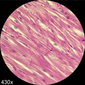
Muscle Tissue: Composed of elongated contractile cells with moderate ECM. Responsible for strong contraction and body movements.
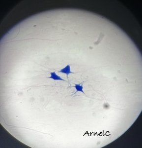
Nervous Tissue: Features elongated cells with extremely fine processes and very little ECM. Specialized for transmission of nerve impulses.
Tissue | Cells | ECM | Functions |
|---|---|---|---|
Epithelium | Aggregated polyhedral cells | Small amount | Lining surfaces, glandular secretion |
Connective | Fixed and wandering cells | Abundant | Support, protection |
Muscle | Elongated contractile cells | Moderate | Contraction, movement |
Nervous | Elongated cells with fine processes | Very small | Transmission of impulses |
Epithelium: Structure and Function
Structural/Functional Forms
Epithelium exists in two main forms:
Sheets of contiguous cells: Cover external and internal surfaces (epithelium).
Clusters of invaginated cells: Form glands (glandular epithelium).
Functional Classification
Covering (surface) epithelium: Lines outer surfaces and mucous/serous membranes of organs.
Glandular epithelium: Specialized for secretion; includes endocrine (hormones) and exocrine (enzymes, sweat) glands.
Functions of Epithelium
Protection: Shields underlying tissues from abrasion and injury.
Transcellular transport: Moves molecules across epithelial sheets.
Secretion: Produces mucinogen, hormones, enzymes, and other molecules.
Absorption: Takes up materials from lumens (e.g., intestines, kidney tubules).
Selective permeability: Controls movement of materials between compartments.
Sensation detection: Specialized cells detect taste, vision, and sound.
Junctional Complexes in Epithelial Cells
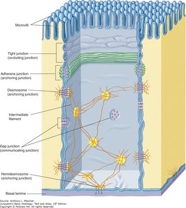
Epithelial cells are tightly bound by specialized junctions:
Tight junctions (zonula occludens): Prevent passive flow between cells.
Adherens junctions (zonula adherens): Stabilize and strengthen cell connections.
Desmosomes: Provide strong attachment points.
Gap junctions: Allow intercellular communication.
Hemidesmosomes: Anchor cells to the basal lamina.

Epithelium: Classification
By Layers (Cell Arrangement)
Simple epithelium: Single layer of cells.
Stratified epithelium: Multiple layers of cells.
Pseudostratified epithelium: Appears multilayered but all cells contact the basal lamina.
Transitional epithelium: Specialized for stretching (e.g., urinary bladder).
By Cell Shape (Morphology)
Squamous: Thin, flat cells.
Cuboidal: Box-like, square cells.
Columnar: Tall, rectangular cells.
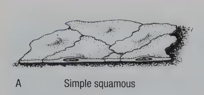
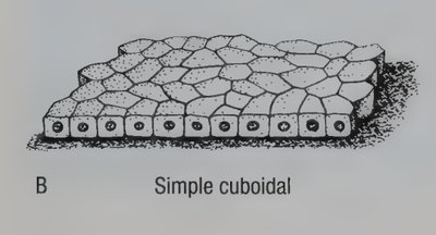
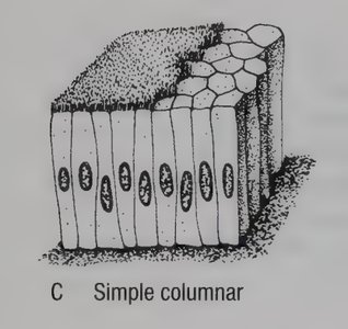
Simple Epithelium
Simple squamous epithelium: Single layer of flat cells; found in alveoli of lungs, lining blood vessels.
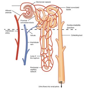
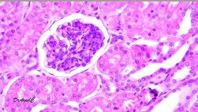
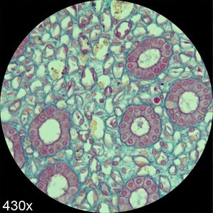
Simple cuboidal epithelium: Single layer of cube-shaped cells; found in kidney tubules, glandular ducts.
Simple columnar epithelium: Single layer of tall, rectangular cells; found in digestive tract lining.
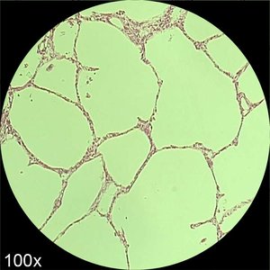
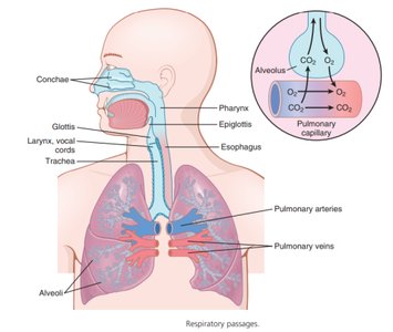
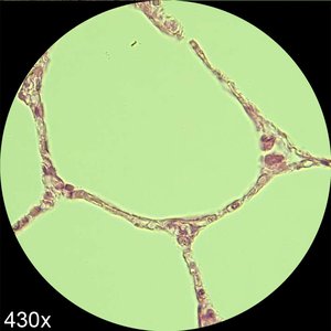
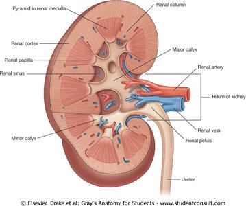
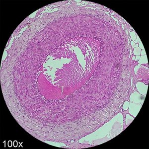
Stratified Epithelium
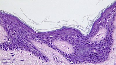
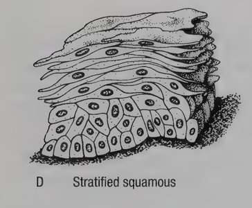
Stratified squamous epithelium: Multiple layers; basal cells are rounded, uppermost cells are flat. Found in skin, oral cavity, esophagus.
Keratinized: Surface cells contain keratin (skin).
Non-keratinized: Surface cells lack keratin (oral cavity, esophagus).
Transitional Epithelium
Transitional epithelium is specialized for stretching and is found in the urinary bladder. Cells change shape from rounded to flat as the bladder fills.
Pseudostratified Epithelium
Pseudostratified epithelium appears multilayered but all cells contact the basal lamina. Commonly found in the respiratory tract, often with cilia.
Glandular Epithelium
Types of Glands
Exocrine glands: Secrete products into ducts (e.g., sweat, sebaceous glands).
Endocrine glands: Secrete hormones directly into the bloodstream (e.g., pancreas, thyroid).
Modes of Secretion
Holocrine: Entire cell disintegrates to release secretion.
Merocrine: Secretion released by exocytosis.
Apocrine: Portion of cell membrane buds off to release secretion.
Classification of Multicellular Exocrine Glands
Multicellular exocrine glands are classified based on duct structure and secretory unit shape:
Simple glands: Unbranched ducts.
Compound glands: Branched ducts.
Tubular: Tube-shaped secretory units.
Acinar: Round, sac-like secretory units.
Summary Table: Epithelial Tissue Classification
Type | Layers | Shape | Location | Function |
|---|---|---|---|---|
Simple Squamous | 1 | Flat | Alveoli, blood vessels | Diffusion, filtration |
Simple Cuboidal | 1 | Cube | Kidney tubules, glands | Secretion, absorption |
Simple Columnar | 1 | Tall | Digestive tract | Absorption, secretion |
Stratified Squamous | Multiple | Flat (surface) | Skin, oral cavity | Protection |
Transitional | Multiple | Variable | Urinary bladder | Stretching |
Pseudostratified | Appears multiple | Tall | Respiratory tract | Secretion, movement |
Key Terms and Concepts
Extracellular Matrix (ECM): Non-cellular component providing structural support.
Basal Lamina: Thin layer separating epithelium from connective tissue.
Keratinization: Process by which cells become filled with keratin protein.
Connexons: Protein complexes forming gap junctions.
Equations and Scientific Notation
While histology does not typically involve equations, cell surface area and volume can be calculated for comparative purposes:
Surface Area of a Cell:
Volume of a Cell:
Additional info:
Some images and diagrams were inferred to represent classic histological examples based on context and standard academic knowledge.
Tables and classification schemes were expanded for clarity and completeness.
