 뒤로
뒤로Study Guide: Tissues & The Integumentary System (Anatomy & Physiology)
스터디 가이드 - 스마트 노트
 자료에 맞춘 맞춤형 노트, 핵심 정의, 예시, 맥락을 확장해 제공합니다.
Q1. Briefly define tissue.
Background
Topic: Tissue Definition
This question is testing your understanding of what constitutes a tissue in the context of human anatomy and physiology.
Key Terms:
Tissue: A group of cells that work together to perform a specific function.
Cells: The basic structural and functional units of life.
Step-by-Step Guidance
Recall that tissues are collections of similar cells.
Think about how these cells are organized and what they do collectively.
Consider the role of tissues in forming organs and organ systems.
Try solving on your own before revealing the answer!
Final Answer:
A tissue is a group of similar cells that are organized to carry out a specific function in the body.
This organization allows tissues to form organs and contribute to overall physiological processes.
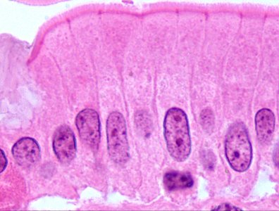
Q2. What are hematoxylin and eosin? ii. In the figure below, identify the cellular structures stained by each.
Background
Topic: Histology Stains
This question is testing your knowledge of common stains used in histology and your ability to identify which cellular structures are stained by hematoxylin and eosin.
Key Terms:
Hematoxylin: A basic dye that stains acidic structures (like nuclei) a deep blue or purple.
Eosin: An acidic dye that stains basic structures (like cytoplasm and extracellular matrix) pink.
Histology: The study of tissues under the microscope.
Step-by-Step Guidance
Recall what hematoxylin and eosin are used for in tissue staining.
Think about which cellular structures are acidic (e.g., nuclei) and which are basic (e.g., cytoplasm).
Examine the provided image and note the color differences between the nuclei and the surrounding cytoplasm.
Identify which structures are stained purple/blue and which are stained pink.
Try solving on your own before revealing the answer!
Final Answer:
Hematoxylin stains the nuclei (which appear purple/blue), while eosin stains the cytoplasm and extracellular matrix (which appear pink).
In the image, the dark purple oval structures are nuclei stained by hematoxylin, and the lighter pink areas are cytoplasm stained by eosin.
Q3. Identify and describe the various features of the 4 classes of tissue: Epithelial, Connective, Muscular, Nervous
Background
Topic: Tissue Classification
This question is testing your ability to distinguish between the four main tissue types in the human body and describe their features.
Key Terms:
Epithelial Tissue: Covers surfaces, lines cavities, forms glands.
Connective Tissue: Supports, binds, and protects other tissues and organs.
Muscular Tissue: Responsible for movement.
Nervous Tissue: Conducts electrical impulses and processes information.
Step-by-Step Guidance
List the four tissue types: epithelial, connective, muscular, and nervous.
For each type, describe its main function (e.g., protection, support, movement, communication).
Consider cellularity: Which tissues have tightly packed cells (epithelial, muscular, nervous) and which have more extracellular matrix (connective)?
Think about subcategories (e.g., epithelial: squamous, cuboidal, columnar; connective: bone, blood, cartilage).
Sketch or visualize an example for each tissue type and note typical locations in the body.
Reflect on regenerative capability: Which tissues regenerate well (epithelial, connective) and which do not (nervous)?
Try solving on your own before revealing the answer!
Final Answer:
Epithelial: Covers surfaces, lines cavities, forms glands; tightly packed cells; subcategories include squamous, cuboidal, columnar; found in skin, lining of organs; high regenerative capability.
Connective: Supports, binds, protects; fewer cells, more extracellular matrix; subcategories include bone, blood, cartilage; found throughout body; variable regenerative capability.
Muscular: Movement; elongated cells; subcategories include skeletal, cardiac, smooth; found in muscles; moderate regenerative capability.
Nervous: Communication; highly specialized cells (neurons); found in brain, spinal cord, nerves; low regenerative capability.
Each tissue type has distinct features, functions, and regenerative abilities.
Q4. In the figure, label A and B.
Background
Topic: Histology Identification
This question is testing your ability to identify and label structures in a histology image, specifically those stained by hematoxylin and eosin.
Key Terms:
Nucleus: The cell's control center, stained purple/blue by hematoxylin.
Cytoplasm: The cell's internal fluid, stained pink by eosin.
Step-by-Step Guidance
Examine the image and locate the structures labeled A and B.
Identify which structure is stained dark purple/blue (likely the nucleus) and which is stained pink (likely the cytoplasm).
Recall the staining properties of hematoxylin and eosin to help with identification.
Label A and B based on their color and location within the cell.
Try solving on your own before revealing the answer!
Final Answer:
A: Nucleus (stained by hematoxylin, appears purple/blue)
B: Cytoplasm (stained by eosin, appears pink)
The nucleus is the dark oval structure, and the cytoplasm is the lighter area surrounding it.
