 뒤로
뒤로Synapses and Signal Transduction in the Nervous System
스터디 가이드 - 스마트 노트
 자료에 맞춘 맞춤형 노트, 핵심 정의, 예시, 맥락을 확장해 제공합니다.
Action Potentials and Neuronal Signaling
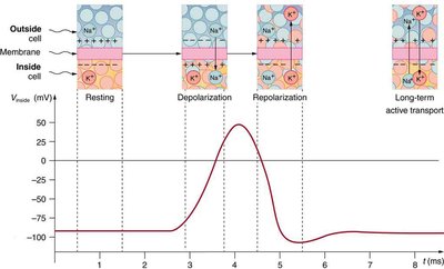
Generation and Propagation of Action Potentials
An action potential is a rapid, temporary change in a cell's membrane potential that allows for the transmission of electrical signals along neurons. This process is fundamental for communication within the nervous system.
Threshold: The minimum membrane potential required to initiate an action potential.
Depolarization: Rapid influx of Na+ ions causes the membrane potential to become more positive.
Repolarization: K+ ions exit the cell, returning the membrane potential toward its resting value.
Refractory Periods: The absolute refractory period prevents another action potential from occurring, while the relative refractory period allows a stronger stimulus to trigger another action potential.
Graded Potentials and Summation
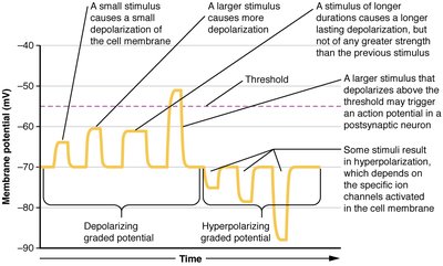
Graded potentials are changes in membrane potential that vary in size and can add together to influence whether a neuron reaches threshold to fire an action potential.
Depolarizing graded potentials move the membrane potential closer to threshold.
Hyperpolarizing graded potentials move the membrane potential further from threshold.
Summation: Temporal summation occurs when signals arrive close together in time; spatial summation occurs when signals arrive from different locations.
Neuronal Structure and Signal Transmission
Neuron Anatomy and Signal Flow
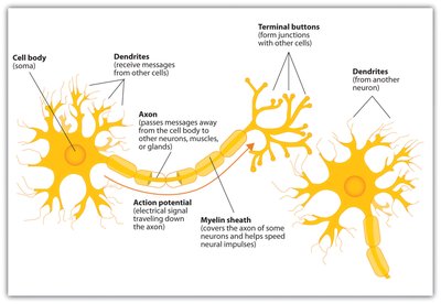
Neurons are specialized cells that transmit electrical and chemical signals. Their structure is adapted for efficient communication.
Dendrites: Receive incoming signals from other neurons.
Cell body (soma): Integrates incoming signals.
Axon: Conducts action potentials away from the cell body.
Myelin sheath: Insulates the axon, increasing conduction velocity via saltatory conduction.
Axon terminals: Release neurotransmitters to communicate with other cells.
Synaptic Transmission
Chemical Synapses
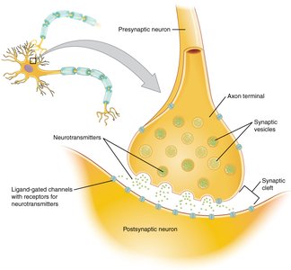
A synapse is the junction where one neuron communicates with another cell. Chemical synaptic transmission involves the release of neurotransmitters that bind to receptors on the postsynaptic cell, altering its membrane potential.
Action potential arrives at the axon terminal.
Voltage-gated Ca2+ channels open, allowing Ca2+ influx.
Neurotransmitter-containing vesicles fuse with the membrane and release their contents into the synaptic cleft.
Neurotransmitters bind to receptors on the postsynaptic cell, causing ion channels to open or close and generating a graded potential.
Cell Communication and Signal Transduction
Types of Chemical Signaling
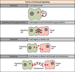
Cells communicate using chemical signals that can act on the same cell, nearby cells, or distant cells. The specificity of the response depends on the presence of appropriate receptors.
Type | Description |
|---|---|
Autocrine | Signal acts on the cell that released it |
Paracrine | Signal acts on nearby cells |
Endocrine | Signal travels through the blood to distant cells |
Gap junction | Direct communication between adjacent cells |
Signal Transduction Pathways
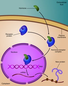
Signal transduction is the process by which a cell converts an external signal into a functional response. This can involve rapid changes in protein activity or slower changes in gene expression and protein synthesis.
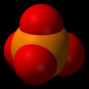
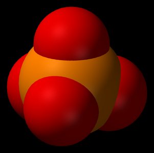
Fast response: Modification of existing proteins, often by phosphorylation (addition of phosphate groups by kinases or removal by phosphatases).
Slow response: Changes in gene expression, leading to altered protein synthesis and longer-lasting effects.
Speed vs. Persistence of Cellular Responses
Cells can respond to signals rapidly by modifying existing proteins or more slowly by changing the abundance of proteins. Fast responses are typically short-lived, while changes in protein population are more persistent.
Change protein activity: Seconds to minutes; uses proteins already present; usually short-lived.
Change protein population: Minutes to hours or longer; requires altered protein production; effects are often more persistent.
Summary
Electrical signals (action potentials) allow rapid communication within neurons, while synapses enable information transfer between cells. Chemical signals and receptors determine the specificity of cellular responses, and signal transduction pathways convert these signals into functional changes, either by modifying existing proteins or altering gene expression.
