 뒤로
뒤로The Cardiovascular System: Cardiac Muscle and Conduction
스터디 가이드 - 스마트 노트
 자료에 맞춘 맞춤형 노트, 핵심 정의, 예시, 맥락을 확장해 제공합니다.
The Cardiovascular System: Cardiac Muscle and Conduction
Cardiac Muscle Structure and Properties
The heart is composed of specialized muscle cells called cardiomyocytes, which are responsible for the contractile function of the heart. These cells have unique structural and functional properties that distinguish them from skeletal muscle cells.
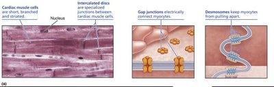
Striated Appearance: Cardiac muscle cells are striated due to the organized arrangement of actin and myosin filaments.
Cell Shape: Cardiomyocytes are short, branched, and typically contain a single nucleus.
Intercalated Discs: Specialized junctions called intercalated discs connect adjacent cardiomyocytes, allowing for synchronized contraction.
Mechanical and Electrical Coupling: Cell-to-cell contact- intercalated discs contain desmosomes (mechanical junctions) and gap junctions (electrical junctions) that facilitate the transmission of force and electrical signals between cells.
Energy Requirements of Cardiac Muscle
Cardiac muscle requires a significant amount of energy to sustain continuous contractions throughout life.
ATP Production: Cardiomyocytes produce and consume approximately 5 kg of ATP per day, relying almost exclusively on aerobic respiration. Cardiomyocytes make ATP almost exclusively from aerobic respiration
Mitochondria: These cells contain exceptionally large and numerous mitochondria to meet their high energy demands.
Oxygen Supply: Adequate oxygen delivery via coronary circulation is critical for cardiac function.
Comparison: Cardiac Muscle vs. Skeletal Muscle
Cardiac muscle and skeletal muscle share some similarities but also have important differences in structure and function.
Feature | Skeletal Muscle | Cardiac Muscle |
|---|---|---|
Structure | Striated, long, cylindrical, multinucleate | Striated, short, branched, one or two nuclei per cell |
Gap Junctions | No | Yes |
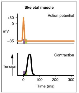
Contraction as a Unit | No, motor units must be stimulated individually | Yes, gap junctions create a functional syncytium |
T tubules | Abundant | Fewer, wider |
Sarcoplasmic Reticulum | Elaborate; has terminal cisterns | Less elaborate; no terminal cisterns |
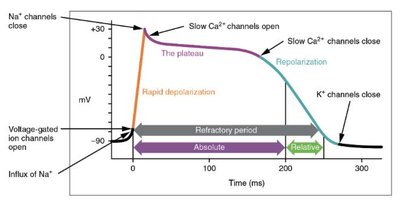
Source of Ca2+ for Contraction | Sarcoplasmic reticulum only | Sarcoplasmic reticulum and extracellular fluid |
Pacemaker Cells | No | Yes |
Tetanus Possible | Yes | No |
ATP Supply | Aerobic and anaerobic (fewer mitochondria) | Aerobic only (more mitochondria) |
Cardiac Conduction System
Overview of the Cardiac Conduction System
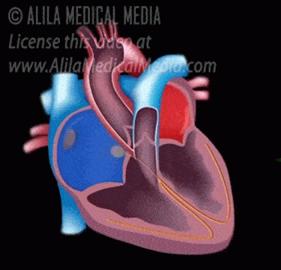
The heart is autorhythmic, meaning it can generate its own electrical impulses to coordinate contraction. This is achieved through a specialized conduction system composed of non-contractile cardiomyocytes.
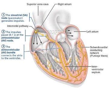
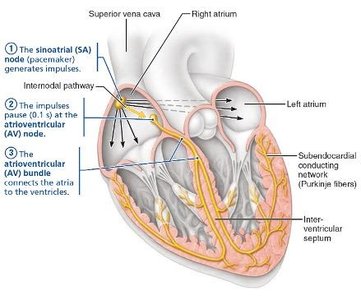
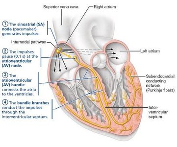
Sinoatrial (SA) Node: The primary pacemaker of the heart, located in the right atrium.
Atrioventricular (AV) Node: Receives impulses from the SA node and delays transmission to allow atrial contraction before ventricular contraction.
AV Bundle (Bundle of His), Bundle Branches, and Purkinje Fibers: Distribute the electrical signal throughout the ventricles.
Steps of the Cardiac Conduction System
SA Node Depolarization: The SA node spontaneously depolarizes, initiating the heartbeat (sinus rhythm, ~75 beats/minute).
Atrial Contraction: The impulse spreads across the atria via gap junctions, causing atrial contraction.
AV Node Activation: The signal reaches the AV node, which delays the impulse to allow complete atrial contraction and ventricular filling.
Impulse Transmission to Ventricles: The signal travels down the AV bundle, bundle branches, and Purkinje fibers, resulting in coordinated ventricular contraction.
Autonomic Regulation of Heart Rate
Although the heart has its own pacemaker, the autonomic nervous system modulates heart rate and force of contraction.
Sympathetic Stimulation: Increases heart rate and contractility via cardiac nerves to the SA and AV nodes and myocardium.
Parasympathetic Stimulation: Decreases heart rate via the vagus nerves.
Electrocardiogram (ECG or EKG)
Principles of ECG
An electrocardiogram (ECG) records the electrical activity of the heart using electrodes placed on the skin. It provides a non-invasive assessment of heart function and can help diagnose various cardiac conditions.
P Wave: Represents atrial depolarization.
QRS Complex: Represents ventricular depolarization (and atrial repolarization, which is masked).
T Wave: Represents ventricular repolarization.
Interpretation of ECG Waves
P Wave: Depolarization of the atria; initiates atrial contraction. The flatline after the P wave is due to the AV node delay, allowing the atria to finish contracting and ventricles to fill.
QRS Complex: Depolarization of the ventricles; the largest electrical event due to the large muscle mass. Atrial repolarization occurs simultaneously but is hidden by the QRS complex.
T Wave: Repolarization of the ventricles, preparing them for the next cycle.
The Cardiac Cycle
Phases of the Cardiac Cycle
The cardiac cycle consists of one complete contraction and relaxation of all four heart chambers. The two main phases are systole (contraction) and diastole (relaxation).
Phase 1 (Ventricular Filling): All chambers are relaxed, AV valves are open, and blood passively fills the ventricles.
Phase 2 (Atrial Systole): The SA node fires, causing atrial contraction and forcing the last 30% of blood into the ventricles (P wave on ECG).
Phase 3 (Isovolumetric Contraction): The AV node fires (QRS complex on ECG), atria relax, ventricles contract, AV valves close, and semilunar valves remain closed.
Phase 4 (Ventricular Ejection): When ventricular pressure exceeds aortic/pulmonary trunk pressure, semilunar valves open and blood is ejected (stroke volume).
Phase 5 (Isovolumetric Relaxation): Ventricles repolarize and relax (T wave on ECG), semilunar valves close, and the cycle repeats as AV valves reopen.
Cardiac Output
Cardiac output (CO) is the volume of blood ejected by each ventricle per minute. It is a key measure of heart function and is calculated as the product of stroke volume (SV) and heart rate (HR):
Stroke Volume (SV): The volume of blood ejected by the ventricles in a single heartbeat (ml/beat).
Heart Rate (HR): The number of heartbeats per minute (beats/min).
Example: If stroke volume is 70 ml/beat and heart rate is 75 beats/min, then cardiac output is:
