 뒤로
뒤로The Cardiovascular System: Structure and Function
스터디 가이드 - 스마트 노트
 자료에 맞춘 맞춤형 노트, 핵심 정의, 예시, 맥락을 확장해 제공합니다.
The Cardiovascular System
Overview
The cardiovascular system is essential for transporting blood, nutrients, gases, and wastes throughout the body. It consists of the heart and a network of blood vessels, forming two main circuits: the pulmonary and systemic circuits.
Heart: A muscular pump that keeps blood circulating through the vessels.
Blood Vessels: Tubular structures (arteries, veins, capillaries) that deliver blood to and from tissues.
Pulmonary and Systemic Circuits
Major Divisions
The cardiovascular system is divided into two main circuits, each serving a distinct function:
Pulmonary Circuit: Carries deoxygenated blood from the right side of the heart to the lungs for gas exchange, then returns oxygenated blood to the left side of the heart.
Systemic Circuit: Delivers oxygenated blood from the left side of the heart to the body tissues and returns deoxygenated blood to the right side of the heart.
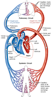
Pulmonary Circuit
The pulmonary circuit is supplied by the right side of the heart. It sends blood to the lungs via the pulmonary arteries and returns oxygenated blood through the pulmonary veins.
Arteries: Carry blood away from the heart (in this circuit, to the lungs).
Veins: Return blood toward the heart (from the lungs).
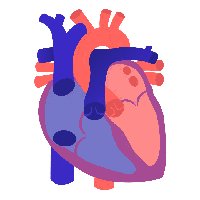
Systemic Circuit
The systemic circuit is supplied by the left side of the heart. It pumps blood into the aorta, the largest artery, and distributes oxygenated blood to all body tissues. Deoxygenated blood returns via the superior and inferior vena cava.
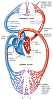
Heart Anatomy
Position, Size, and Shape
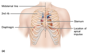
The heart is located in the thoracic cavity, specifically in the mediastinum, and is tilted slightly to the left. The average adult heart weighs about 300 grams.
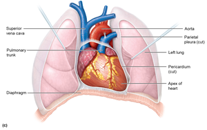
External Landmarks
The base of the heart is the broad superior portion, while the apex is the inferior, pointed end. The apical impulse can be palpated between the fifth and sixth ribs, just below the left nipple.
The Pericardium
Structure and Function
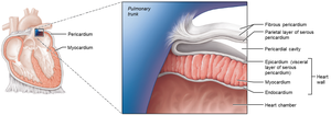
The heart is enclosed in a double-walled sac called the pericardium, which protects and anchors the heart, prevents overfilling, and reduces friction.
Parietal Pericardium: The outer layer.
Visceral Pericardium (Epicardium): The inner layer, directly covering the heart.
Pericarditis
Pericarditis is inflammation of the pericardium, often due to viral infection. It can cause the membranes to become rough, producing a painful friction rub with each heartbeat.
The Heart Wall
Layers of the Heart Wall
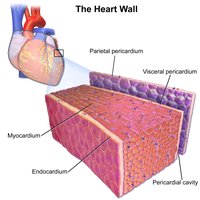
The heart wall consists of three layers, each with distinct functions:
Epicardium: The outermost layer (also the visceral pericardium).
Myocardium: The thick, muscular middle layer responsible for contraction.
Endocardium: The smooth inner lining of the heart chambers and valves.
Heart Chambers
Internal Features
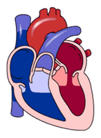
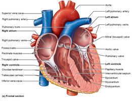
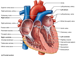
The heart contains four chambers: two superior atria and two inferior ventricles. The interatrial septum separates the atria, while the interventricular septum separates the ventricles. The fossa ovalis is a remnant of the fetal foramen ovale.
Atria
The atria are thin-walled chambers that receive blood returning to the heart from large veins. Auricles are appendages that increase atrial volume. Three systemic veins empty into the right atrium.
Ventricles
The ventricles are thick-walled chambers that receive blood from the atria and eject it into the arteries. The right ventricle forms most of the anterior surface, while the left ventricle dominates the posterior-inferior surface. Key internal features include trabeculae carneae, papillary muscles, and chordae tendineae.
Right Ventricle: Pumps blood into the pulmonary trunk (pulmonary circuit).
Left Ventricle: Pumps blood into the aorta (systemic circuit).
Heart Valves
Types of Valves
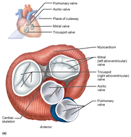
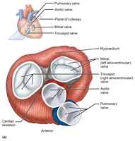
The heart contains four valves, each consisting of 2-3 flaps (cusps) of thin tissue. These valves ensure unidirectional blood flow through the heart.
Atrioventricular (AV) Valves
The AV valves are located between the atria and ventricles. The right AV valve is the tricuspid valve, and the left AV valve is the mitral (bicuspid) valve. Each cusp is anchored to papillary muscles by chordae tendineae, which prevent valve prolapse during ventricular contraction.

Semilunar Valves
Semilunar valves are located between the ventricles and the great arteries. The pulmonary semilunar valve is at the base of the pulmonary trunk, and the aortic semilunar valve is at the base of the aorta. Each has three cusps and prevents backflow into the ventricles.
Blood Flow Through the Heart
Pathway of Blood
Blood flows through the heart in a specific sequence, ensuring separation of oxygenated and deoxygenated blood. Blood in the left and right chambers does not mix, maintaining efficient oxygen delivery to tissues.
Deoxygenated blood enters the right atrium from the body, passes to the right ventricle, and is pumped to the lungs.
Oxygenated blood returns from the lungs to the left atrium, passes to the left ventricle, and is pumped to the body.
Coronary Circulation
Arterial Supply
The heart has its own blood supply, known as coronary circulation. The left coronary artery supplies the left atrium and branches, while the right coronary artery supplies the right atrium and branches. This system ensures the heart receives sufficient oxygen and nutrients to sustain its workload.
Venous Drainage
After passing through the coronary arteries and capillaries, blood drains into veins that empty into the coronary sinus, which returns blood directly to the right atrium.
Clinical Considerations
Homeostatic Imbalances
Blockage of the coronary arteries can lead to serious conditions:
Angina Pectoris: Chest pain due to reduced blood flow to the heart muscle.
Myocardial Infarction (Heart Attack): Death of heart muscle tissue due to prolonged lack of blood supply.
Atherosclerosis
Atherosclerosis is a condition where arteries become stiff and narrowed due to plaque buildup. It begins with vessel damage, followed by accumulation of cholesterol, fat, and connective tissue. Platelets adhere to plaques, stimulating further growth and potentially blocking blood flow. Treatments include bypass surgery, angioplasty, or stent placement.
Condition | Description | Treatment |
|---|---|---|
Angina Pectoris | Chest pain from reduced blood flow | Medications, lifestyle changes |
Myocardial Infarction | Heart muscle death from blocked artery | Emergency intervention, surgery |
Atherosclerosis | Arterial plaque buildup | Bypass, angioplasty, stents |
