뒤로
뒤로The Cardiovascular System: The Heart – Structure, Function, and Circulation
스터디 가이드 - 스마트 노트
 자료에 맞춘 맞춤형 노트, 핵심 정의, 예시, 맥락을 확장해 제공합니다.
Heart Anatomy and Blood Flow
The Systemic and Pulmonary Circuits
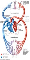
The heart functions as a double pump, supporting two major circulatory pathways: the pulmonary and systemic circuits. Each side of the heart supplies its own circuit, ensuring efficient oxygenation and nutrient delivery throughout the body.
Right Side: Receives deoxygenated blood from body tissues and pumps it to the lungs for oxygenation (pulmonary circuit).
Left Side: Receives oxygenated blood from the lungs and pumps it to the rest of the body (systemic circuit).
Receiving Chambers (Atria): Right atrium receives blood from the systemic circuit; left atrium receives blood from the pulmonary circuit.
Pumping Chambers (Ventricles): Right ventricle pumps blood into the pulmonary circuit; left ventricle pumps blood into the systemic circuit.
Size, Location, and Orientation of the Heart
Gross Anatomy and Position
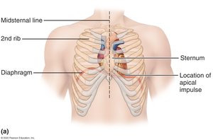
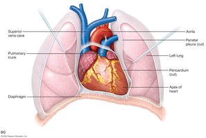
The heart is approximately the size of a fist and weighs less than 1 pound. It is located in the mediastinum, superior to the diaphragm, with about two-thirds of its mass to the left of the midsternal line. The apex points toward the left hip, and the heart is positioned anterior to the vertebral column and posterior to the sternum.
Structure of the Heart Wall
Three Layers of the Heart Wall
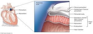
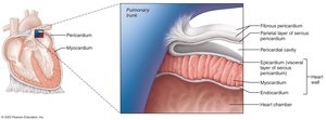
The heart wall is composed of three distinct layers, each with specialized structure and function:
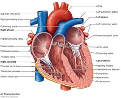
Epicardium: The visceral layer of the serous pericardium, forming the outermost layer.
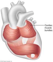
Myocardium: Consists of circular or spiral bundles of contractile cardiac muscle cells, responsible for the heart's pumping action.
Endocardium: The innermost layer, continuous with the endothelial lining of blood vessels, lining the heart chambers and covering the cardiac skeleton of valves.
Chambers and Associated Great Vessels
Heart Chambers
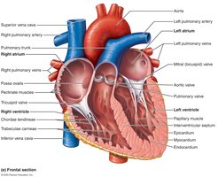
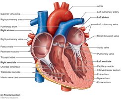
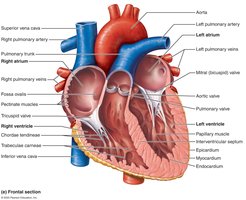
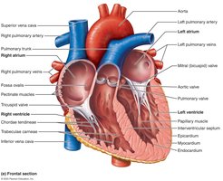
The heart contains four chambers: two superior atria and two inferior ventricles. The interventricular septum separates the right and left ventricles.
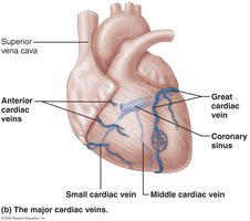
Atria: Small, thin-walled receiving chambers. The right atrium receives deoxygenated blood from the superior and inferior vena cava and the coronary sinus. The left atrium receives oxygenated blood from the four pulmonary veins.
Ventricles: Thick-walled discharging chambers. The right ventricle pumps blood into the pulmonary trunk, while the left ventricle pumps blood into the aorta, the largest artery in the body.
Papillary Muscles: Anchor the chordae tendineae, which are attached to the heart valves.
Heart Valves
Types and Functions of Heart Valves
Heart valves ensure unidirectional blood flow through the heart, opening and closing in response to pressure changes. There are two main types:
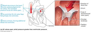
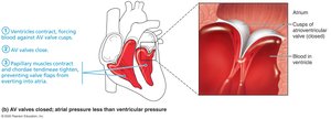
Atrioventricular (AV) Valves: Located between the atria and ventricles (tricuspid on the right, mitral on the left).
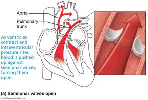
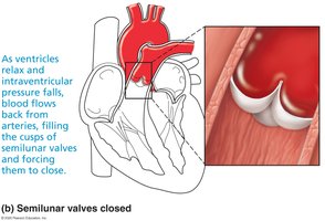
Semilunar (SL) Valves: Located between the ventricles and major arteries (pulmonary and aortic valves).
Function of the Atrioventricular (AV) Valves
AV valves open when atrial pressure exceeds ventricular pressure, allowing blood to flow from atria to ventricles.
When ventricles contract, the increased pressure forces AV valves to close, preventing backflow into the atria.
Function of the Semilunar (SL) Valves
SL valves open when ventricular pressure rises above arterial pressure, allowing blood to be ejected into the arteries.
When ventricles relax, arterial blood fills the cusps of the SL valves, causing them to close and preventing backflow into the ventricles.
Clinical Considerations: Valve Disorders
Incompetent Valve: Blood backflows, causing the heart to repump the same blood repeatedly.
Valvular Stenosis: Stiff valve flaps constrict the opening, requiring the heart to exert more force to pump blood.
Treatment: Valve replacement may be necessary in severe cases.
Pathway of Blood Through the Heart
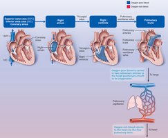
Right Side (Pulmonary Circuit)
Superior vena cava (SVC), inferior vena cava (IVC), and coronary sinus →
Right atrium →
Tricuspid valve →
Right ventricle →
Pulmonary semilunar valve →
Pulmonary trunk →
Pulmonary arteries →
Lungs
Left Side (Systemic Circuit)
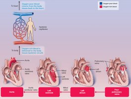
Four pulmonary veins →
Left atrium →
Mitral valve →
Left ventricle →
Aortic semilunar valve →
Aorta →
Systemic circulation
Comparative Features of Pulmonary and Systemic Circuits
Equal volumes of blood are pumped to both circuits.
Pulmonary circuit is short and low-pressure; systemic circuit is long and high-friction.
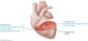
The left ventricle has walls three times thicker than the right ventricle, reflecting the greater force needed for systemic circulation.
Coronary Circulation
Blood Supply to the Heart Muscle
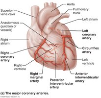
Coronary circulation provides the heart muscle (myocardium) with its own blood supply. This is the shortest circulation in the body and is delivered primarily when the heart is relaxed. The left ventricle receives the majority of the coronary blood supply.