 뒤로
뒤로The Cell Cycle and Mitosis: Structure, Phases, and Microscopic Identification
스터디 가이드 - 스마트 노트
 자료에 맞춘 맞춤형 노트, 핵심 정의, 예시, 맥락을 확장해 제공합니다.
Chapter 3: Cell Cycle & Mitosis
Overview of the Cell Cycle
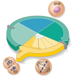
The cell cycle describes the sequence of events that a cell undergoes from its formation until it divides into two daughter cells. It is essential for growth, tissue repair, and asexual reproduction in multicellular organisms. The cell cycle consists of two major phases: Interphase and the Mitotic (M) phase.
Interphase: The period of cell growth and normal function, including DNA replication and preparation for division.
Mitotic (M) phase: The period during which the cell divides its nucleus (mitosis) and cytoplasm (cytokinesis).
Interphase
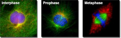
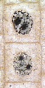
Interphase is the longest phase of the cell cycle, during which the cell grows, performs its normal functions, and prepares for division. The nuclear material is in the form of chromatin. Interphase is subdivided into three subphases:
G1 phase (Gap 1): Vigorous cell growth and metabolism occur. The cell increases in size and synthesizes proteins and organelles.
S phase (Synthesis): DNA replication takes place, ensuring that each daughter cell will receive an identical set of chromosomes.
G2 phase (Gap 2): The cell continues to grow and prepares for mitosis by synthesizing proteins and organelles required for cell division.
Checkpoints in the Cell Cycle
Checkpoints are control mechanisms that ensure the cell is ready to proceed to the next stage. The most important are the G1 checkpoint (restriction point) and the G2 checkpoint, which verify DNA integrity and readiness for division.
Mitosis: Division of the Nucleus
Mitosis is the process by which a eukaryotic cell separates its duplicated chromosomes into two identical sets, resulting in two genetically identical daughter nuclei. Mitosis is divided into four main stages: Prophase, Metaphase, Anaphase, and Telophase. Cytokinesis, the division of the cytoplasm, usually overlaps with telophase.
Prophase
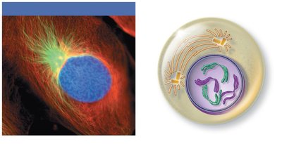
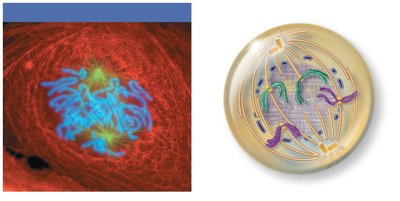
During prophase, chromatin condenses into visible chromosomes, each consisting of two sister chromatids joined at a centromere. The mitotic spindle begins to form as centrosomes move to opposite poles of the cell. The nuclear envelope starts to break down in late prophase, and spindle fibers attach to kinetochores on the chromosomes.
Metaphase
In metaphase, the chromosomes align at the cell's equator, known as the metaphase plate. This alignment ensures that each daughter cell will receive one copy of each chromosome.
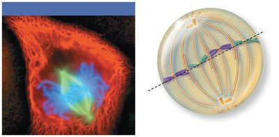
Anaphase
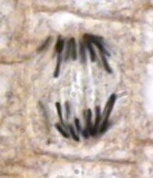
Anaphase is the shortest phase of mitosis. The centromeres split, and the sister chromatids (now individual chromosomes) are pulled toward opposite poles of the cell by the spindle fibers. This separation ensures equal distribution of genetic material.
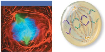
Telophase
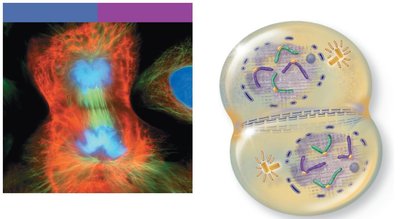
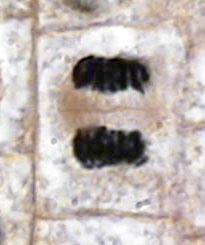
During telophase, the chromosomes reach the poles and begin to decondense back into chromatin. New nuclear envelopes form around each set of chromosomes, and nucleoli reappear. The spindle apparatus disassembles.
Cytokinesis
Cytokinesis is the division of the cytoplasm, which usually begins during late anaphase or telophase. A contractile ring of actin filaments forms a cleavage furrow that pinches the cell into two daughter cells, each with a nucleus identical to the original.
Microscopic Identification of Mitosis Stages
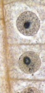
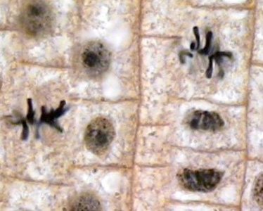
Microscopic examination of cells, such as those in an Allium (onion) root tip, allows for the identification of different mitotic stages based on chromosomal appearance and cell structure.
Stage | Key Features | Representative Image |
|---|---|---|
Interphase | Nucleus visible, chromatin not condensed |
|
Prophase | Chromosomes condense, nuclear envelope breaks down |
|
Metaphase | Chromosomes aligned at metaphase plate |
|
Anaphase | Chromatids separate and move to poles |
|
Telophase/Cytokinesis | Chromosomes decondense, nuclear envelope reforms, cell divides |
|
Summary Table: Mitosis Stages and Key Events
Stage | Main Events |
|---|---|
Prophase | Chromosomes condense, spindle forms, nuclear envelope breaks down |
Metaphase | Chromosomes align at metaphase plate |
Anaphase | Sister chromatids separate and move to opposite poles |
Telophase | Chromosomes decondense, nuclear envelope reforms |
Cytokinesis | Cytoplasm divides, two daughter cells form |
Key Terms and Definitions
Chromatin: The uncondensed form of DNA and protein found in the nucleus during interphase.
Chromosome: A structure consisting of DNA and proteins that becomes visible during mitosis.
Centromere: The region where two sister chromatids are joined together.
Mitotic spindle: A structure made of microtubules that separates chromosomes during mitosis.
Kinetochore: A protein structure on the centromere where spindle fibers attach.
Cleavage furrow: The indentation that begins the process of cytokinesis in animal cells.
Example: Application in Tissue Growth
Mitosis is crucial for the growth of multicellular organisms, repair of damaged tissues, and replacement of old or dead cells. For example, the rapid division of cells in the root tip of an onion (Allium) allows the root to grow and penetrate the soil.
Additional info: The cell cycle is tightly regulated by cyclins and cyclin-dependent kinases (CDKs), which ensure that each phase is completed accurately before the next begins. Errors in cell cycle regulation can lead to uncontrolled cell division and cancer.
