 뒤로
뒤로The Cellular Level of Organization: Structure and Function of Cells
스터디 가이드 - 스마트 노트
 자료에 맞춘 맞춤형 노트, 핵심 정의, 예시, 맥락을 확장해 제공합니다.
The Cellular Level of Organization
Introduction to Cells
Cells are the smallest living units in the human body and serve as the fundamental building blocks of all tissues and organs. Each cell maintains homeostasis, and the collective activity of cells ensures the stability of tissues, organs, and the entire organism.
Sex cells (germ cells or reproductive cells): Sperm in males and oocytes in females, responsible for reproduction.
Somatic cells: All other body cells except sex cells.
Plasma Membrane
Structure and Function
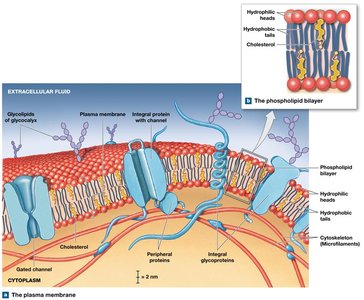
The plasma membrane forms the outer boundary of the cell, providing selective transport and communication with the environment. Its main components are lipids, proteins, and carbohydrates.
Physical isolation: Separates the cytoplasm from the extracellular fluid.
Regulation of exchange: Controls entry and exit of ions, nutrients, and wastes.
Sensitivity: Contains receptors for environmental signals.
Structural support: Anchors cells and stabilizes tissues.
Membrane Lipids
Phospholipid bilayer: Hydrophilic heads face outward; hydrophobic tails form the core, acting as a barrier to ions and water-soluble compounds.
Cholesterol: Reduces membrane fluidity and permeability.
Membrane Proteins
Integral proteins: Embedded within the membrane; some span the entire membrane (transmembrane proteins).
Peripheral proteins: Bound to the inner or outer surface.
Functional types:
Recognition proteins: Identify cells as normal or abnormal.
Receptor proteins: Bind ligands (e.g., hormones).
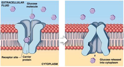
Carrier proteins: Transport specific solutes.
Channels: Permit water and small solutes to pass.
Membrane Carbohydrates
Glycocalyx: A "sugar coat" formed by proteoglycans, glycoproteins, and glycolipids.
Functions: Lubrication, protection, specificity in binding, and cell recognition (immune response).
Transport Across the Plasma Membrane
Types of Transport
Passive transport: No energy required (e.g., diffusion, osmosis).
Active transport: Requires energy (ATP).
Carrier-mediated transport: Can be passive or active.
Vesicular transport: Always active (requires ATP).
Diffusion
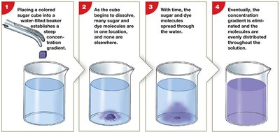
Diffusion is the net movement of a substance from an area of higher concentration to an area of lower concentration, proceeding down a concentration gradient until equilibrium is reached.
Factors affecting diffusion rate: Distance, molecule size, temperature, and gradient size.
Diffusion Across Plasma Membranes
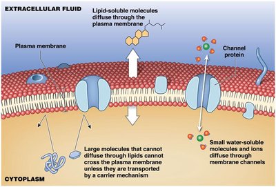
Simple diffusion: Lipid-soluble compounds and dissolved gases cross the lipid bilayer.
Channel-mediated diffusion: Water, ions, and small molecules pass through protein channels.
Osmosis
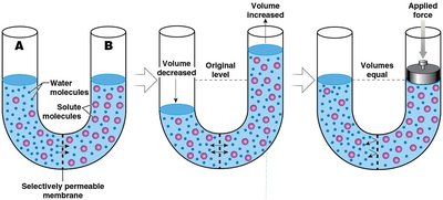
Osmosis is the net diffusion of water across a selectively permeable membrane toward a higher solute concentration. It continues until solute concentrations are equal on both sides.
Osmotic pressure: The force with which pure water moves into a solution due to solute concentration.
Hydrostatic pressure: Opposes osmotic pressure and prevents further osmosis.
Aquaporins: Water channels that facilitate rapid osmosis.
Tonicity
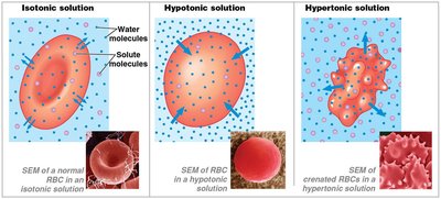
Tonicity describes how the concentration of solutes in a solution affects cell volume.
Solution Type | Solute Concentration | Effect on Cell |
|---|---|---|
Isotonic | Equal to cell | No net water movement; cell remains unchanged |
Hypotonic | Lower than cell | Water enters cell; cell may swell and burst (hemolysis) |
Hypertonic | Higher than cell | Water leaves cell; cell shrinks (crenation) |
Carrier-Mediated and Vesicular Transport
Carrier-Mediated Transport
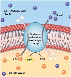
Active transport: Moves substances against their concentration gradients using ATP (e.g., ion pumps, sodium-potassium pump).
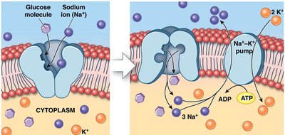
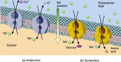
Secondary active transport: Uses gradients established by primary active transport to move other substances without direct ATP use.
Symport (cotransport): Two substances move in the same direction.
Antiport (countertransport): Two substances move in opposite directions.
Facilitated diffusion: Passive transport of large or insoluble molecules via carrier proteins.
Vesicular Transport (Bulk Transport)
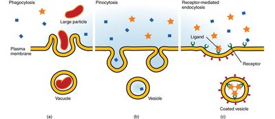
Endocytosis: Imports extracellular materials via vesicles.
Types of endocytosis:
Receptor-mediated endocytosis: Specific ligands are brought into the cell.
Pinocytosis: Uptake of extracellular fluid.
Phagocytosis: Uptake of solid particles by pseudopodia.
Exocytosis: Release of substances from the cell as vesicles fuse with the plasma membrane.
Cellular Organelles
Overview
Organelles are specialized structures within the cytoplasm that perform specific functions necessary for cell survival and activity.
Cytoplasm: All material between the plasma membrane and the nucleus.
Cytosol: Intracellular fluid containing water, nutrients, ions, proteins, and waste products.
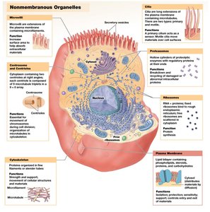
Nonmembranous organelles: Not enclosed by a membrane (e.g., cytoskeleton, centrioles, ribosomes, microvilli, cilia, flagella).
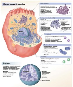
Membranous organelles: Surrounded by a membrane (e.g., endoplasmic reticulum, Golgi apparatus, lysosomes, peroxisomes, mitochondria).
Nonmembranous Organelles
Cytoskeleton: Protein framework for shape, strength, and flexibility.
Microvilli: Increase surface area for absorption.
Centrioles: Form spindle apparatus during cell division.
Cilia: Move fluids or secretions across cell surface.
Flagellum: Enables sperm cell movement.
Ribosomes: Sites of protein synthesis.
Membranous Organelles
Smooth Endoplasmic Reticulum (SER): Synthesizes phospholipids, cholesterol, steroid hormones, and stores glycogen and triglycerides.
Rough Endoplasmic Reticulum (RER): Studded with ribosomes; synthesizes proteins.
Golgi Apparatus: Modifies, packages, and delivers secretory products; renews plasma membrane; packages enzymes in lysosomes.
Lysosomes: Vesicles containing digestive enzymes; responsible for autolysis (self-destruction of damaged cells).
Peroxisomes: Break down fatty acids and neutralize hydrogen peroxide with catalase.
Mitochondria: Produce ATP via aerobic metabolism (cellular respiration).
The Nucleus
Structure and Function
Nuclear envelope: Double membrane surrounding the nucleus.
Nuclear pores: Allow chemical communication between nucleus and cytoplasm.
Nucleoplasm: Fluid inside the nucleus.
Nucleolus: Synthesizes rRNA and assembles ribosomal subunits.
The nucleus stores genetic information (DNA) and controls protein synthesis. Genes are sequences of DNA that code for proteins, dictating cell structure and function.
Protein Synthesis
Transcription
Transcription is the synthesis of RNA from a DNA template. Messenger RNA (mRNA) carries genetic instructions from the nucleus to the cytoplasm for protein synthesis.
Translation
Translation is the synthesis of a polypeptide based on mRNA instructions. Ribosomes read mRNA codons, and transfer RNA (tRNA) delivers amino acids to build the protein.
Genetic Control
Mutations: Permanent changes in DNA that can alter protein structure and function.
The Cell Life Cycle
Cell Division
Cell division produces two identical daughter cells for growth and tissue repair. The cell life cycle includes interphase (G1, S, G2 phases) and the M phase (mitosis and cytokinesis).
Interphase: Cell grows, duplicates organelles, and replicates DNA.
Mitosis: Division of the nucleus (prophase, metaphase, anaphase, telophase).
Cytokinesis: Division of the cytoplasm, resulting in two daughter cells.
Apoptosis: Programmed cell death.
Regulation of the Cell Cycle
Stimulation: Internal factors (M-phase promoting factor, MPF) and extracellular growth factors.
Inhibition: Repressor genes and short telomeres.
Cell Division and Cancer
Tumor (neoplasm): Mass of abnormal cell growth.
Benign tumor: Remains localized.
Malignant tumor: Invades surrounding tissues and can metastasize.
Cancer: Caused by mutations in genes regulating cell growth and division (oncogenes); mutagens and carcinogens are contributing factors.
Cellular Differentiation
All somatic cells contain the same genetic material, but differentiation occurs by turning off specific genes, allowing cells to become specialized (e.g., liver cells, neurons).
