 뒤로
뒤로The Cellular Level of Organization: Structure and Function of Cells
스터디 가이드 - 스마트 노트
 자료에 맞춘 맞춤형 노트, 핵심 정의, 예시, 맥락을 확장해 제공합니다.
The Cellular Level of Organization
Introduction
The cell is the fundamental unit of structure and function in all living organisms. Understanding the anatomy and physiology of cells is essential for comprehending how the human body operates at the most basic level. This chapter explores the structure, components, and transport mechanisms of cells.
Plasma Membrane
Structure and Function
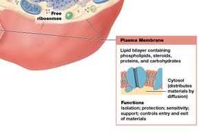
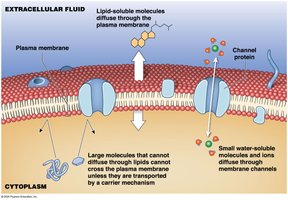
The plasma membrane (cell membrane) forms the outer boundary of the cell and regulates the movement of substances into and out of the cell. It is primarily composed of a lipid bilayer with embedded proteins.
Main Components: Lipids (mainly phospholipids), proteins, and carbohydrates.
Functions: Isolation, protection, sensitivity, support, and control of entry and exit of materials.
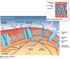
Types of Membrane Proteins by Function
Receptor proteins: Bind and respond to ligands (e.g., ions, hormones).
Carrier proteins: Bind and transport specific solutes across the membrane.
Channels: Integral proteins with a central pore for water and small solutes; may be gated to regulate passage.
Cellular Organelles
Nonmembranous Organelles
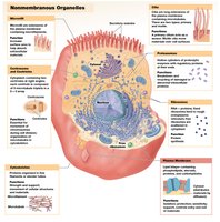
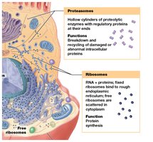
Nonmembranous organelles are not surrounded by a lipid membrane and are found throughout the cytoplasm.
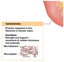
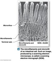
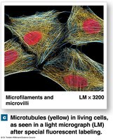
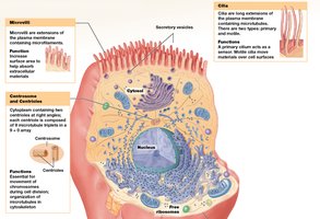
Cytoskeleton: Provides structural support, shape, and movement for the cell.
Microvilli: Increase surface area for absorption.
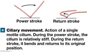
Cilia: Move fluids or secretions across the cell surface; primary cilia are sensory, motile cilia are for movement.
Centrioles: Organize microtubules during cell division.
Proteasomes: Break down and recycle damaged or abnormal proteins.
Ribosomes: Synthesize proteins; can be free in cytoplasm or bound to rough ER.
Membranous Organelles
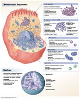
Membranous organelles are surrounded by lipid membranes, compartmentalizing their functions within the cell.
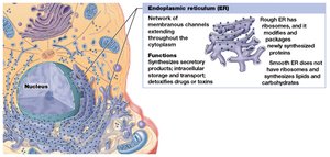
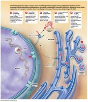
Endoplasmic Reticulum (ER): Network of membranes; rough ER synthesizes proteins, smooth ER synthesizes lipids and detoxifies chemicals.
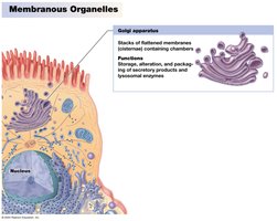
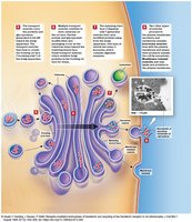
Golgi Apparatus: Modifies, sorts, and packages proteins and lipids for secretion or delivery to other organelles.
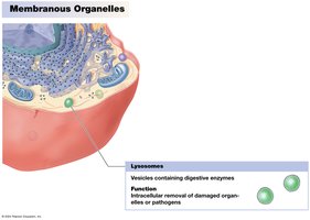
Lysosomes: Contain digestive enzymes for intracellular removal of damaged organelles or pathogens.
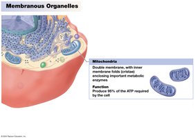
Mitochondria: Produce ATP, the cell’s main energy currency, through aerobic respiration.
The Nucleus
Structure and Function
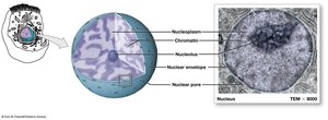
The nucleus is the largest organelle and serves as the control center for cellular operations.
Controls cellular metabolism
Stores and processes genetic information (DNA)
Controls protein synthesis
Transport Across the Plasma Membrane
Types of Transport
Passive Transport: Does not require energy (e.g., diffusion, osmosis).
Active Transport: Requires energy (ATP) to move substances against their concentration gradients.
Carrier-Mediated Transport: Can be passive (facilitated diffusion) or active.
Vesicular Transport: Always active; involves movement of materials in vesicles (endocytosis, exocytosis).
Diffusion
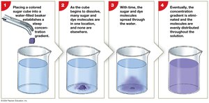
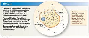
Diffusion is the net movement of a substance from an area of higher concentration to an area of lower concentration, down its concentration gradient.
Concentration Gradient: The difference in concentration between two areas.
Diffusion continues until equilibrium is reached, but molecular motion persists.
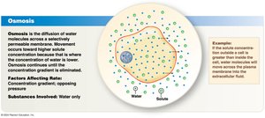
Osmosis
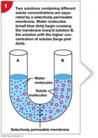
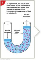
Osmosis is the net diffusion of water across a selectively permeable membrane toward the solution with higher solute concentration.
Water moves to balance solute concentrations on both sides of the membrane.
Osmotic Pressure: The pressure required to stop the osmotic flow of water.
Tonicity
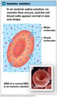
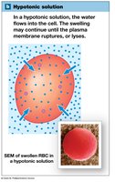
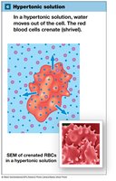
Tonicity describes how the concentration of solutes in a solution affects cell volume.
Isotonic Solution: Equal solute concentration; no net water movement; cell remains unchanged.
Hypotonic Solution: Lower solute concentration outside; water enters cell; cell may swell and burst (hemolysis).
Hypertonic Solution: Higher solute concentration outside; water leaves cell; cell shrinks (crenation).
Carrier-Mediated and Vesicular Transport
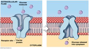
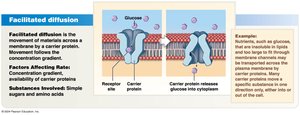
Facilitated Diffusion
Facilitated diffusion is the passive movement of molecules across the membrane via specific carrier proteins. It is used for substances that are too large or insoluble in lipids.
Examples: Glucose, amino acids
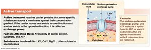
Active Transport
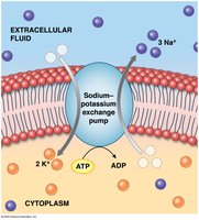
Active transport uses energy (usually ATP) to move substances against their concentration gradients. Ion pumps are a key example.
Primary Active Transport: Direct use of ATP (e.g., sodium–potassium exchange pump).
Sodium–Potassium Exchange Pump: Moves 3 Na+ out and 2 K+ in per ATP hydrolyzed.
Secondary Active Transport
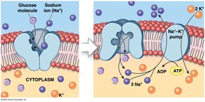
Secondary active transport uses the energy from the movement of one substance down its gradient to drive the transport of another substance against its gradient.
Example: Glucose transport coupled with sodium ions.
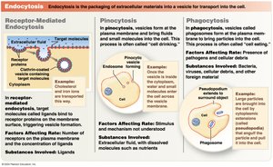
Vesicular Transport
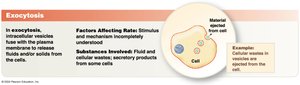
Vesicular (bulk) transport involves the movement of large particles or volumes of fluid into or out of the cell via vesicles. This process requires ATP.
Endocytosis: Import of materials into the cell (includes phagocytosis and pinocytosis).
Exocytosis: Export of materials out of the cell.
The Membrane Potential
Definition and Importance
Membrane potential is the electrical potential difference across the plasma membrane, resulting from the unequal distribution of positive and negative charges. It is essential for nerve impulse transmission and muscle contraction.
Resting membrane potential in unstimulated cells ranges from −10 mV to −100 mV (inside more negative than outside).
Summary Table: Types of Membrane Transport
Transport Type | Energy Required? | Direction | Example Substances |
|---|---|---|---|
Simple Diffusion | No | Down gradient | O2, CO2 |
Facilitated Diffusion | No | Down gradient | Glucose, amino acids |
Osmosis | No | Down water gradient | Water |
Active Transport | Yes (ATP) | Against gradient | Na+, K+ |
Vesicular Transport | Yes (ATP) | Bulk movement | Proteins, bacteria |
