 뒤로
뒤로The Circulatory System: Blood – Structure, Function, and Clinical Relevance
스터디 가이드 - 스마트 노트
 자료에 맞춘 맞춤형 노트, 핵심 정의, 예시, 맥락을 확장해 제공합니다.
The Circulatory System: Blood
Introduction
The circulatory system is essential for transporting substances throughout the body and maintaining homeostasis. Blood, as a connective tissue, plays a central role in this system, working alongside the heart and blood vessels to support life.
The circulatory system consists of the heart, blood vessels, and blood.
Not to be confused with the cardiovascular system (heart and blood vessels only)
Functions of Blood in the Circulatory System
Transport: Blood carries oxygen, nutrients, hormones, and waste products to and from cells.
Protection: Blood contains immune cells (white blood cells) and proteins that defend against pathogens and initiate clotting to prevent blood loss.
Pathogens are neutralized and destroyed
Platelets in the blood initiate clotting
Regulation: Blood helps regulate body temperature, pH, and fluid balance.
By buffering acids and bases, the blood maintains a stable pH
Routes blood outwards (give off heat), or inwards (keep heat)
Components and General Properties of Blood
Volume: 4–6 liters in adults, about 8% of body weight.
Men have more blood content than women.
Temperature: Approximately 38°C (100.4°F).
Viscosity: About 5 times more viscous than water, important for flow and resistance.
Water is 5x thicker than water! This slows down the speed of fluid movement through the capillaries so that oxygen, hormones, and nutrients had time to diffuse out of the blood vessels.
Shear stress in blood vessels is the frictional force of flowing blood applied parallel against the inner lining of the blood vessel walls.
Blood can become too thick when you're dehydrated, causing an increase in blood pressure and forcing the heart to work harder.
pH: 7.35–7.45 (slightly alkaline).
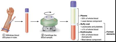
Blood is composed of plasma (the liquid matrix) and formed elements (red and white blood cells and cell fragments/platelets).
Blood Plasma (55%)
Plasma is the clear yellowish, liquid portion of blood, making up about 55% of total blood volume. It is primarily water but contains important solutes:
Water (92%)
Proteins (7%): Albumin, globulins, and fibrinogen
Other solutes (1%): Nutrients, wastes, hormones, gases, electrolytes
Plasma is essential for:
Clotting
Immune defense
Transport of iron, lipids, and hydrophobic hormones
Plasma Proteins (all sourced from the liver)
Albumin: Most abundant; maintains osmotic pressure, transports substances, and buffers pH of plasma.
Majorly contributes to the thickness (viscosity) of blood and the blood osmolarity. Blood osmolarity is the concentration of particles that can pass through blood vessels.
Globulins: Involved in immune defense and transport of lipids and fat-soluble vitamins.
Immunity, clotting, and solute transport.
Fibrinogen: Essential for blood clotting.
Soluble precursors to fibrin.
Formed Elements of Blood (45%)
Formed elements include erythrocytes (red blood cells), leukocytes (white blood cells), and platelets. They make up about 45% of blood volume.
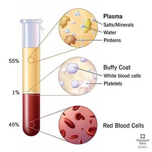
Red blood cells (45%) are the most dense, followed by white blood cells (1%), then plasma (55%)
Table: Main Components of Blood
Component | Percentage | Main Function |
|---|---|---|
Plasma | ~55% | Transport of nutrients, wastes, hormones |
Buffy Coat (WBCs & Platelets) | ~1% | Immune defense, clotting |
Red Blood Cells | ~45% | Oxygen and carbon dioxide transport |
How Blood is Produced (Hematopoiesis)
Blood cells are continuously produced in the red bone marrow through a process called hematopoiesis. Every day, the body produces billions of new blood cells to replace old or lost ones lost through urination.
Hematopoiesis: Production of formed elements
Erythropoiesis: Production of red blood cells
200 billion/day
Leukopoiesis: Production of white blood cells
10 billion/day
Thrombopoiesis: Production of platelets
400 billion/day
Erythrocytes (Red Blood Cells)
Quantity and Structure
Most abundant formed element in blood responsible for picking up oxygen from the lungs and transporting it to the tissues.
Red blood cells transport oxygen by using a specialized iron-rich protein called hemoglobin to bind and carry oxygen from the lungs to body tissues.
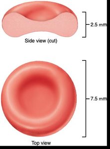
Biconcave disc shape: Increases surface area for gas exchange
Lack a nucleus and most organelles
This means they cannot divide or repair themselves)
No nucleus = (1) more surface area for carrying oxygen, (2) less turbulence for movement through small capillaries.
Life span = 120 days
Hemoglobin
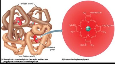
Hemoglobin is a red, iron-containing protein that binds and transports oxygen and carbon dioxide. Each hemoglobin molecule can carry up to four oxygen molecules.
Composed of four polypeptide chains (globins) and four heme groups
Iron in heme binds oxygen. One iron can bind to one oxygen molecule.
One hemoglobin protein can carry up to four oxygen molecules.
RBC lack mitochondria so they must rely on anaerobic fermentation to meet ATP needs.
Red Blood Cell Life Cycle
Produced in red bone marrow from hematopoietic stem cells through erythropoiesis. They originate from hematopoietic stem cells,
Live about 120 days. They are born and die at roughly the same rate. It takes 3-4 days to produce mature RBCs.
Old RBCs are removed by the spleen and liver; components are recycled
Sickle-cell Anemia: Sickle cells occur due to genetic disorder where a single amino acid (valine) is changed in the beta chain of the hemoglobin. It is a recessive gene (two copies of the HbS gene). Carriers with one copy do not typically present symptoms of the disorders.
Treatments: Gene therapy
Benefits: cannot get malaria if two copies of the gene are present.
Blood Types
Blood types are determined by the presence or absence of specific antigens on the surface of red blood cells. The main blood group systems are ABO and Rh.
ABO System: Types A, B, AB, and O, based on A and B antigens
Rh System: Presence (+) or absence (−) of Rh antigen (D antigen)
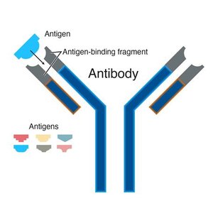
Table: ABO Blood Types
Blood Type | Antigens on RBCs | Antibodies in Plasma |
|---|---|---|
A | A | Anti-B |
B | B | Anti-A |
AB | A and B | None |
O | None | Anti-A and Anti-B |
Leukocytes (White Blood Cells)
Structure and Function
Leukocytes are immune cells that protect the body from infection and foreign substances. They are classified as granulocytes or agranulocytes based on the presence of granules in their cytoplasm and reside primarily in the cytoplasm.
Granulocytes: Neutrophils, eosinophils, basophils
Agranulocytes: Lymphocytes, monocytes
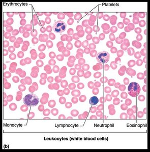
Table: Types of Leukocytes
Type | Main Function |
|---|---|
Neutrophils (Granulocyte) |
|
Eosinophils (Granulocyte) |
|
Basophils (Granulocyte) |
|
Lymphocytes (Agranulocyte) |
|
Monocytes (Agranulocyte) |
|
Leukocyte Life Cycle
Leukopoiesis: Leukocytes are produced in the bone marrow from hematopoietic stem cells just like RBC. Leukocytes do NOT stay in the red bone marrow.
Their lifespan varies from hours to years, depending on the type and immune activity.
Disorders
Leukemias: group of cancerous conditions involving overproduction of abnormal WBCs involving clones of single abnormal cell.
Infectious mononucleosis: highly contagious viral disease caused by the Epstein-Barr virus.
usually seen in young adults
results in excess lymphocytes
symptoms: tired, achy, sore throat, low grade fever persisting 4-6 weeks
Platelets and Control of Bleeding
Platelet Structure and Function
Small fragments of bone marrow cells known as megakaryocytes
Second most abundant formed element
Essential for blood clotting (hemostasis)
Control of Bleeding (Hemostasis)
Hemostasis is the process of stopping blood loss from damaged vessels. It involves three main stages:
Vascular spasm: The smooth muscle contracts, constricting the of blood vessel to reduce blood flow (vasoconstriction).
Platelet plug formation: Platelets adhere to exposed collagen and aggregate. The platelets release chemicals that make the nearby platelets sticky and allow a plug to form.
Coagulation: Fibrin forms a mesh that traps blood cells, forming a stable clot
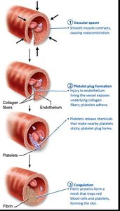
Agglutination vs. Coagulation
Coagulation: Formation of a blood clot via fibrin meshwork.
Agglutination: Clumping of red blood cells due to antibody-antigen interactions (important in blood typing)
This occurs when there is a poor blood type match. Antibodies cause the blood cells to stick together.
Blood Transfusions and Typing
Transfusing Red Blood Cells
Whole-blood transfusions are used in cases of rapid, substantial blood loss.
Packed red blood cells with plasma and white blood cells removed (PRBCs) are preferred for restoring oxygen-carrying capacity.
PRBCs have a shelf life of up to 42 days
Blood must be matched for ABO and Rh types to prevent fatal reactions.
Blood Typing: Universal Donor and Recipient
Based on interactions between
Antigens: cell surface marks
How our body determines its own cells from foreign invaders:
Antibodies: recognize and bind to antigens to initiate an immune response
When antibodies bind to antigens on cells, causes them to stick together
Ex. Type A has Anti-B antibodies that will react with type B blood upon exposure.
Agglutination: For blood, body naturally produces antibodies against other blood types antigens
Universal donor: Type O (no antigens).
Can donate to all blood types, but can only receive from self.
Universal recipient: Type AB (no antibodies)
Can receive donations from all blood types but can only donate to self.
Antibodies are present in plasma, not in transfused red blood cells
Rh Factor
Rh antigen is either present (+) or absent (−)
Rh-negative individuals can develop antibodies if exposed to Rh-positive blood
Important consideration in pregnancy (hemolytic disease of the newborn)
Summary Table: Blood Components and Functions
Component | Main Function |
|---|---|
Plasma | Transport, osmotic balance, pH buffering |
Erythrocytes | Oxygen and carbon dioxide transport |
Leukocytes | Immune defense |
Platelets | Blood clotting |
