 뒤로
뒤로The Endocrine System: Structure, Function, and Regulation
스터디 가이드 - 스마트 노트
 자료에 맞춘 맞춤형 노트, 핵심 정의, 예시, 맥락을 확장해 제공합니다.
The Endocrine System
Overview of the Endocrine System
The endocrine system is a major regulatory system of the body, responsible for the production and secretion of hormones that regulate various physiological processes. It works closely with the nervous system to maintain homeostasis, growth, metabolism, and reproduction.
Hormones are chemical messengers secreted by endocrine glands into the bloodstream, affecting distant target organs.
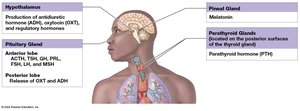
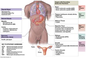
The endocrine system includes glands such as the pituitary, thyroid, parathyroid, adrenal, pineal, and pancreas, as well as organs with secondary endocrine functions (e.g., heart, kidneys, gonads).
Intercellular Communication
Mechanisms of Intercellular Communication
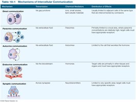
Cells communicate to coordinate activities and maintain homeostasis through several mechanisms:
Mechanism | Transmission | Chemical Mediators | Distribution of Effects |
|---|---|---|---|
Direct communication | Gap junctions | Ions, small solutes, lipid-soluble materials | Limited to adjacent cells of the same type |
Paracrine communication | Extracellular fluid | Paracrine factors | Primarily within a single tissue |
Autocrine communication | Extracellular fluid | Autocrines | Limited to the cell that secretes the hormone |
Endocrine communication | Bloodstream | Hormones | Target cells in distant tissues and organs |
Synaptic communication | Across synapses | Neurotransmitters | Limited to specific area; rapid, short-lived effects |
Endocrine vs. Nervous System Communication
Nervous system: Fast, short-lived responses via neurotransmitters at synapses.
Endocrine system: Slower, longer-lasting effects via hormones in the bloodstream.
Both systems use chemical messengers and negative feedback to regulate homeostasis.
Hormones: Structure, Transport, and Action
Classes of Hormones
Hormones are classified based on their chemical structure:
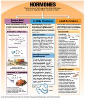
Amino acid derivatives (biogenic amines): Derived from tyrosine (e.g., thyroid hormones, catecholamines) or tryptophan (e.g., melatonin).
Peptide hormones: Chains of amino acids, including glycoproteins (TSH, LH, FSH) and short polypeptides (ADH, OXT, GH, PRL, insulin).
Lipid derivatives: Eicosanoids (from arachidonic acid) and steroid hormones (from cholesterol, e.g., androgens, estrogens, corticosteroids).
Transport and Inactivation of Hormones
Hydrophilic hormones circulate freely and are quickly inactivated.
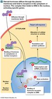
Hydrophobic hormones (thyroid and steroid hormones) bind to transport proteins, creating a reserve in the bloodstream and remaining active longer.
Mechanisms of Hormone Action
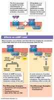
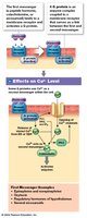
Hormones bind to specific receptors on or in target cells.
Extracellular receptors: For hydrophilic hormones; activate second messenger systems (e.g., cAMP, Ca2+).
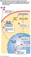
Intracellular receptors: For hydrophobic hormones; directly affect gene expression and protein synthesis.
Regulation of Hormone Secretion
Primarily controlled by negative feedback.
Stimuli include humoral (changes in blood composition), hormonal (other hormones), and neural (neurotransmitter signals).
The Pituitary Gland (Hypophysis)
Anatomy and Control
The pituitary gland is located in the sella turcica, connected to the hypothalamus by the infundibulum. It consists of two lobes:
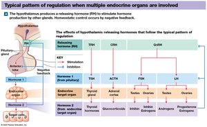
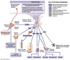
Anterior lobe (adenohypophysis): Produces and releases hormones under hypothalamic control via the hypophyseal portal system.
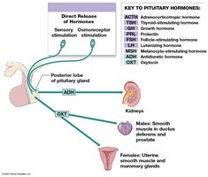
Posterior lobe (neurohypophysis): Stores and releases hormones produced by the hypothalamus (ADH, OXT).
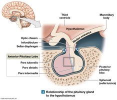
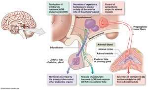
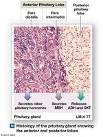
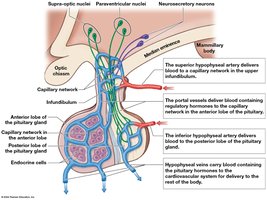
Hormones of the Anterior Lobe
Thyroid-stimulating hormone (TSH): Stimulates thyroid hormone release.
Adrenocorticotropic hormone (ACTH): Stimulates glucocorticoid release from adrenal cortex.
Gonadotropins (FSH, LH): Regulate reproductive organs and hormone production.
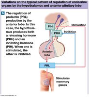
Prolactin (PRL): Stimulates mammary gland development and milk production.
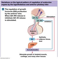
Growth hormone (GH): Stimulates growth, protein synthesis, and metabolism.
Melanocyte-stimulating hormone (MSH): Stimulates melanin production (mainly in fetal development and certain conditions).
Hormones of the Posterior Lobe
Antidiuretic hormone (ADH): Promotes water retention by kidneys; inhibited by alcohol.
Oxytocin (OXT): Stimulates uterine contractions, milk ejection, and sexual arousal.
The Thyroid Gland
Anatomy and Histology
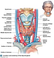
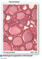
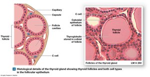
The thyroid gland is located inferior to the thyroid cartilage and consists of two lobes connected by an isthmus. It contains follicles filled with colloid and surrounded by follicular cells, as well as parafollicular (C) cells.
Thyroid Hormones
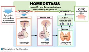
Thyroxine (T4) and Triiodothyronine (T3): Regulate metabolism, growth, and development.
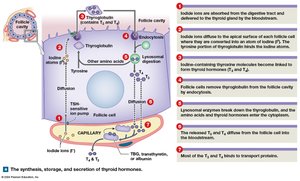
Synthesis: Involves iodide uptake, thyroglobulin production, and enzymatic formation of T3 and T4.
Regulation: Controlled by TSH from the anterior pituitary; transported in blood bound to proteins (TBG, transthyretin, albumin).
Effects of Thyroid Hormones
Increase oxygen consumption and ATP production.
Stimulate metabolism, heat production (calorigenic effect), and development of skeletal, muscular, and nervous systems.
Increase heart rate, blood pressure, and sensitivity to sympathetic stimulation.
Calcitonin
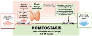
Produced by C cells; lowers blood calcium by increasing excretion by kidneys and decreasing absorption in the digestive tract.
Important during childhood and for reducing bone loss during pregnancy and starvation.
Parathyroid Glands
Anatomy and Function
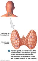
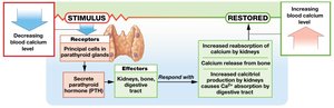
Four small glands on the posterior surface of the thyroid. Principal cells secrete parathyroid hormone (PTH) in response to low blood calcium.
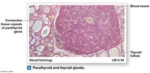
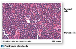
Effects of Parathyroid Hormone (PTH)
Increases blood calcium by stimulating osteoclasts, enhancing kidney reabsorption, and promoting calcitriol synthesis for increased intestinal absorption.
Adrenal Glands
Anatomy and Regions
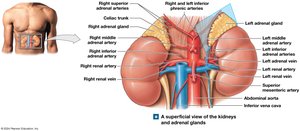
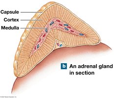
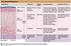
Located superior to each kidney, each adrenal gland consists of an outer cortex and inner medulla.
Adrenal Cortex Hormones
Mineralocorticoids (e.g., aldosterone): Regulate sodium and potassium balance.
Glucocorticoids (e.g., cortisol): Regulate metabolism, stress response, and have anti-inflammatory effects.
Androgens: Minor role in adults; contribute to pubic hair development and muscle formation in females.
Adrenal Medulla Hormones
Epinephrine and norepinephrine: Released in response to sympathetic stimulation; increase heart rate, mobilize energy reserves, and prepare the body for "fight or flight".
Pineal Gland
Location and Function
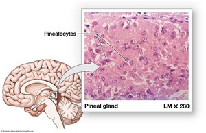
The pineal gland is located in the epithalamus and produces melatonin, which regulates circadian rhythms, inhibits reproductive functions, and acts as an antioxidant.
Pancreas
Anatomy and Function
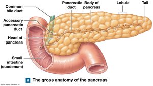
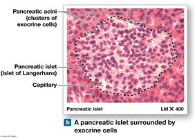
The pancreas is both an exocrine and endocrine gland. The endocrine portion consists of pancreatic islets that secrete hormones regulating blood glucose.
Pancreatic Hormones
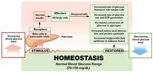
Insulin (from beta cells): Lowers blood glucose by promoting uptake, storage, and utilization of glucose.
Glucagon (from alpha cells): Raises blood glucose by stimulating glycogen breakdown and gluconeogenesis.
Somatostatin (from delta cells): Inhibits insulin and glucagon secretion.
Pancreatic polypeptide (from PP cells): Regulates pancreatic enzyme secretion and gallbladder contraction.
Secondary Endocrine Functions
Other Organs with Endocrine Roles
Kidneys: Produce calcitriol (calcium homeostasis), erythropoietin (RBC production), and renin (blood pressure regulation).
Heart: Produces natriuretic peptides to lower blood pressure and volume.
Thymus: Produces thymosins for immune cell development.
Gonads: Testes produce androgens and inhibin; ovaries produce estrogens, progesterone, and inhibin.
Adipose tissue: Produces leptin, which regulates appetite and reproductive function.
Hormone Interactions and Stress Response
Types of Hormone Interactions
Antagonistic: Opposing effects (e.g., insulin vs. glucagon).
Synergistic: Additive effects (e.g., GH and glucocorticoids).
Permissive: One hormone enables another to act (e.g., thyroid hormone and epinephrine).
Integrative: Different but complementary effects (e.g., calcitriol and PTH).
General Adaptation Syndrome (GAS)
Alarm phase: Immediate, fight-or-flight response (epinephrine dominant).
Resistance phase: Long-term metabolic adjustments (glucocorticoids dominant).
Exhaustion phase: Failure of homeostasis, potentially fatal.
Aging and the Endocrine System
Most hormone levels remain stable with age, but reproductive hormones decline and tissue responsiveness may decrease.
