 뒤로
뒤로The Endocrine System: Structure, Function, and Regulation
스터디 가이드 - 스마트 노트
 자료에 맞춘 맞춤형 노트, 핵심 정의, 예시, 맥락을 확장해 제공합니다.
The Endocrine System: Overview and Major Organs
Introduction to the Endocrine System
The endocrine system is one of the body's two major control systems, working alongside the nervous system to coordinate and integrate the activity of most body cells. It uses chemical messengers called hormones, which are transported in the blood to influence metabolic activities throughout the body. Endocrine responses are typically slower but longer-lasting than nervous system responses.
Endocrinology: The study of hormones and endocrine organs.
Major processes controlled by the endocrine system include reproduction, growth and development, maintenance of electrolyte, water, and nutrient balance, regulation of cellular metabolism, and mobilization of body defenses.
Comparison of Nervous and Endocrine Systems
Feature | Nervous System | Endocrine System |
|---|---|---|
Response Initiation | Rapid | Slow |
Duration | Short | Long |
Signal Type | Action potentials, neurotransmitters | Hormones in blood |
Target Location | Specific (axon pathways) | Diffuse (anywhere blood reaches) |
Distance | Short | Long |
Signal Strength | Frequency of action potentials | Hormone concentration |
Endocrine vs. Exocrine Glands
Exocrine glands: Produce nonhormonal substances (e.g., sweat, saliva) and have ducts to carry secretion to a membrane surface.
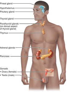
Endocrine glands: Produce hormones, are ductless, and secrete hormones directly into the surrounding extracellular fluid. Major endocrine glands include the pituitary, thyroid, parathyroid, adrenal, and pineal glands. The hypothalamus is a neuroendocrine organ.
Other organs with endocrine tissue: pancreas, gonads, placenta, stomach, intestine, heart, kidneys, skin, thymus, bone, and adipose tissue.
Chemical Nature and Mechanisms of Hormone Action
Hormone Structure and Solubility
The chemical structure of a hormone determines its solubility in water, which affects its transport in blood, degradation rate, and receptor interaction.
Amino acid–based hormones: Most hormones; water-soluble (except thyroxine); cannot cross plasma membrane.
Steroid hormones: Synthesized from cholesterol; lipid-soluble; can cross plasma membrane; includes gonadal and adrenocortical hormones.
Eicosanoids: Sometimes considered hormones, but mostly act as paracrines and autocrines due to localized effects.
Hormone Receptors and Target Cell Specificity
Although hormones circulate to virtually all tissues, only cells with specific receptors for a hormone are affected (target cells). Hormones alter target cell activity by increasing or decreasing the rates of normal cellular processes.
Changes induced by hormones include altering membrane permeability, stimulating protein synthesis, activating/deactivating enzymes, inducing secretory activity, and stimulating mitosis.
Mechanisms of Hormone Action
Water-soluble hormones (all amino acid–based except thyroid hormone): Act on plasma membrane receptors; cannot enter cell; usually act via G protein–coupled second messenger systems.
Lipid-soluble hormones (steroid and thyroid hormones): Act on intracellular receptors that directly activate genes; can diffuse across plasma membrane.
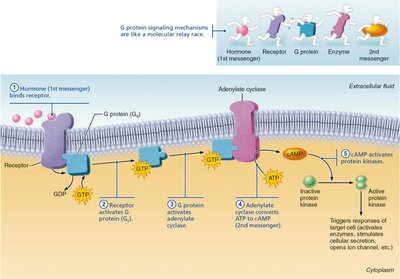
Cyclic AMP (cAMP) Second Messenger System
Hormone (first messenger) binds to receptor.
Receptor activates a G protein.
G protein activates (or inhibits) adenylate cyclase.
Adenylate cyclase converts ATP to cAMP (second messenger).
cAMP activates protein kinases, which phosphorylate other proteins, leading to cellular responses.
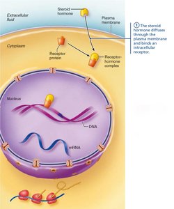
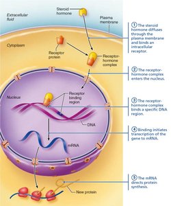
Direct Gene Activation by Lipid-Soluble Hormones
Lipid-soluble hormone diffuses into target cell and binds to intracellular receptor.
Receptor-hormone complex enters nucleus and binds to specific DNA region.
Binding initiates transcription of the gene to mRNA.
mRNA directs protein synthesis.
Regulation of Hormone Release
Types of Endocrine Gland Stimuli
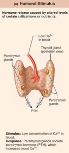
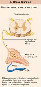
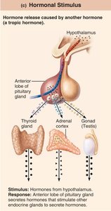
Hormone release is primarily regulated by negative feedback mechanisms and can be triggered by three types of stimuli:
Humoral stimuli: Changing blood levels of ions/nutrients directly stimulate hormone release (e.g., low blood Ca2+ stimulates parathyroid hormone release).
Neural stimuli: Nerve fibers stimulate hormone release (e.g., sympathetic nervous system stimulates adrenal medulla to secrete catecholamines).
Hormonal stimuli: Hormones stimulate other endocrine organs to release their hormones (e.g., hypothalamic hormones regulate anterior pituitary hormones).
Feedback Mechanisms
Negative feedback: Rising hormone levels inhibit further hormone release, maintaining homeostasis.
Nervous system modulation: The nervous system can override or adjust endocrine controls during stress or emergencies.
Pituitary Gland and Hypothalamic Control
Anatomy and Function of the Pituitary Gland
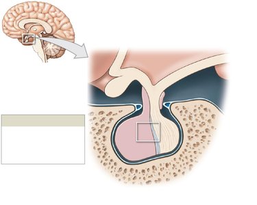
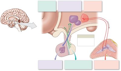
The pituitary gland (hypophysis) is connected to the hypothalamus via the infundibulum and consists of two major lobes:
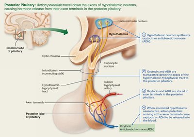
Posterior pituitary (neurohypophysis): Neural tissue; stores and secretes oxytocin and antidiuretic hormone (ADH) produced by the hypothalamus.
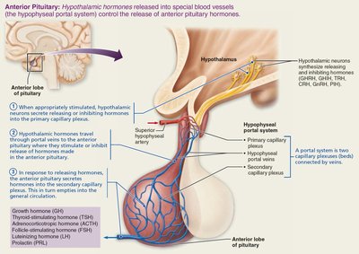
Anterior pituitary (adenohypophysis): Glandular tissue; manufactures and secretes six hormones (GH, TSH, ACTH, FSH, LH, PRL).
Hypothalamic Control of the Pituitary
Posterior pituitary: Hypothalamic neurons synthesize oxytocin and ADH, which are transported down axons and stored in the posterior pituitary. Release is triggered by nerve impulses.
Anterior pituitary: Hypothalamic hormones reach the anterior pituitary via the hypophyseal portal system, regulating hormone secretion.
Major Pituitary Hormones: Regulation and Effects
Posterior Pituitary Hormones
Oxytocin: Stimulates uterine contractions during childbirth and milk ejection during breastfeeding; regulated by positive feedback mechanisms.
Antidiuretic hormone (ADH): Promotes water reabsorption in kidneys; secretion is triggered by high blood osmolarity and inhibited by alcohol.
Anterior Pituitary Hormones
Growth hormone (GH): Stimulates growth (especially bone and muscle) and metabolic functions; regulated by GHRH and GHIH from the hypothalamus.
Thyroid-stimulating hormone (TSH): Stimulates thyroid gland to release thyroid hormones; regulated by TRH from the hypothalamus.
Adrenocorticotropic hormone (ACTH): Stimulates adrenal cortex to release corticosteroids; regulated by CRH from the hypothalamus.
Follicle-stimulating hormone (FSH) and Luteinizing hormone (LH): Regulate function of gonads; secretion triggered by GnRH.
Prolactin (PRL): Stimulates milk production; regulated mainly by PIH (dopamine).
The Thyroid Gland and Its Hormones
Structure and Function
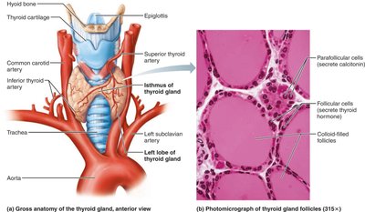
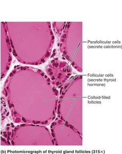
The thyroid gland is a butterfly-shaped organ located on the anterior trachea. It contains follicles that produce thyroglobulin, the precursor to thyroid hormone (TH), and parafollicular cells that produce calcitonin.
Thyroid Hormone (TH)
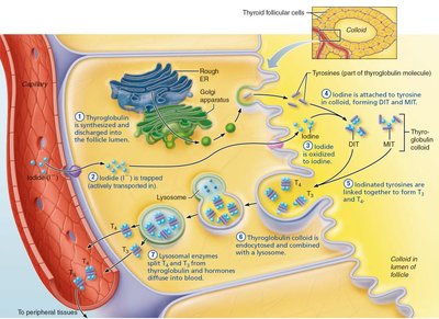
TH exists as thyroxine (T4) and triiodothyronine (T3); both are lipid-soluble and regulate metabolism, growth, and development.
TH increases basal metabolic rate, heat production, and is critical for normal development of the nervous and skeletal systems.
TH synthesis involves iodination of tyrosine residues in thyroglobulin and coupling to form T3 and T4.
Regulation of Thyroid Hormone Secretion
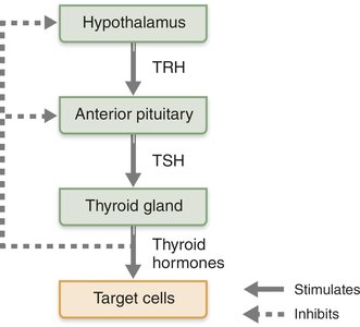
Regulated by negative feedback involving the hypothalamus (TRH), anterior pituitary (TSH), and thyroid gland (TH).
Thyroid Disorders
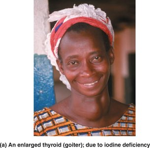
Hypothyroidism: Low TH; symptoms include low metabolic rate, weight gain, and goiter (if due to iodine deficiency).
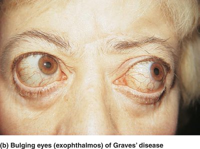
Hyperthyroidism (Graves' disease): High TH; symptoms include high metabolic rate, weight loss, and exophthalmos (bulging eyes).
Calcitonin
Produced by parafollicular (C) cells in response to high blood Ca2+ levels; inhibits osteoclast activity and promotes Ca2+ uptake into bone.
The Parathyroid Glands and Calcium Homeostasis
Structure and Function
The parathyroid glands are small glands located on the posterior aspect of the thyroid gland. They secrete parathyroid hormone (PTH), the primary regulator of blood calcium levels.
PTH increases blood Ca2+ by stimulating osteoclasts, enhancing kidney reabsorption of Ca2+, and activating vitamin D to increase intestinal absorption of Ca2+.
The Adrenal Glands: Structure and Hormones
Adrenal Cortex
Mineralocorticoids (e.g., aldosterone): Regulate Na+ and K+ balance, blood volume, and pressure.
Glucocorticoids (e.g., cortisol): Influence metabolism, help resist stress, and have anti-inflammatory effects.
Gonadocorticoids (e.g., androgens): Contribute to secondary sex characteristics and libido.
Adrenal Medulla
Secretes catecholamines (epinephrine and norepinephrine) during the fight-or-flight response, increasing heart rate, blood pressure, and blood glucose.
The Pancreas and Blood Glucose Regulation
Structure and Function
The pancreas is a mixed gland with both exocrine (digestive enzymes) and endocrine (hormones) functions. The endocrine portion consists of pancreatic islets containing alpha cells (glucagon) and beta cells (insulin).
Glucagon: Raises blood glucose by promoting glycogen breakdown and gluconeogenesis in the liver.
Insulin: Lowers blood glucose by promoting cellular uptake, glycogen synthesis, and inhibiting gluconeogenesis.
Diabetes Mellitus
Type 1: Insulin deficiency due to autoimmune destruction of beta cells.
Type 2: Insulin resistance in target tissues.
Symptoms: Polyuria, polydipsia, polyphagia, and risk of ketoacidosis.
Other Endocrine Organs and Hormones
Pineal gland: Secretes melatonin, regulating sleep-wake cycles.
Gonads: Ovaries produce estrogens and progesterone; testes produce testosterone.
Other tissues: Heart (ANP), kidneys (erythropoietin, renin), adipose tissue (leptin), skeleton (osteocalcin), skin (cholecalciferol), thymus (thymosins).
Developmental and Clinical Aspects
Endocrine glands arise from all three germ layers during development.
Endocrine function generally declines with age, affecting metabolism, growth, and glucose tolerance.
Environmental pollutants can disrupt hormone function, especially sex hormones, thyroid hormone, and glucocorticoids.
