 뒤로
뒤로The Integumentary System: Structure and Function
스터디 가이드 - 스마트 노트
 자료에 맞춘 맞춤형 노트, 핵심 정의, 예시, 맥락을 확장해 제공합니다.
The Integumentary System
Overview and Functions
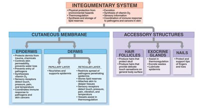
The integumentary system is the body's largest organ system, serving as the primary barrier between the internal environment and the external world. It consists of the skin and its accessory structures, providing protection, sensation, thermoregulation, and metabolic functions.
Physical protection from environmental hazards
Thermoregulation through sweat and blood flow
Synthesis of vitamin D3
Sensory information detection
Excretion of wastes via sweat
Storage of lipid reserves
Cutaneous Membrane
Structure of the Skin
The skin is composed of two main layers: the epidermis (superficial) and the dermis (deep). Beneath the dermis lies the hypodermis (subcutaneous layer), which is not technically part of the integumentary system but provides insulation and energy storage.
Epidermis: Stratified squamous epithelium; avascular; provides protection.
Dermis: Connective tissue; supports and nourishes the epidermis; contains blood vessels, nerves, and accessory structures.
Hypodermis: Adipose tissue; anchors skin to underlying tissues.
Epidermis
Layers of the Epidermis
The epidermis is organized into distinct layers (strata). Thick skin (palms, soles) has five layers; thin skin (most of the body) has four. The layers, from superficial to deep, are:
Stratum corneum
Stratum lucidum (only in thick skin)
Stratum granulosum
Stratum spinosum
Stratum basale (germinativum)
Mnemonic: Come, Let's Get Sun Burned (Corneum, Lucidum, Granulosum, Spinosum, Basale)
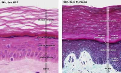
Table: Epidermal Layers and Their Characteristics
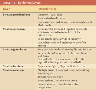
Layer | Characteristics |
|---|---|
Stratum germinativum (basale) | Innermost, basal layer; attached to basal lamina; contains stem cells, melanocytes, and Merkel cells |
Stratum spinosum | Keratinocytes bound by desmosomes; some cell division; Langerhans cells and melanocytes present |
Stratum granulosum | Keratinocytes produce keratohyalin and keratin; cells flatten and die; cell membranes thicken |
Stratum lucidum | Glassy layer; only in thick skin |
Stratum corneum | Multiple layers of dead, flattened cells; water-resistant; slow water loss by insensible perspiration |
Cell Types in the Epidermis
Keratinocytes: Main cell type; produce keratin for strength and waterproofing.
Melanocytes: Located in the stratum basale; produce melanin pigment for UV protection.
Langerhans cells: Immune cells in the stratum spinosum.
Merkel cells: Sensory receptors for light touch in the stratum basale.
Thick vs. Thin Skin
Thick skin: Five layers; found on palms and soles; no hair follicles or sebaceous glands.
Thin skin: Four layers; covers most of the body; contains hair follicles and sebaceous glands.
Dermis
Layers of the Dermis
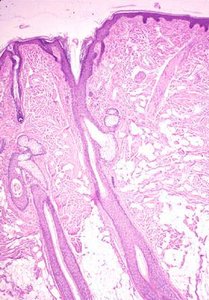
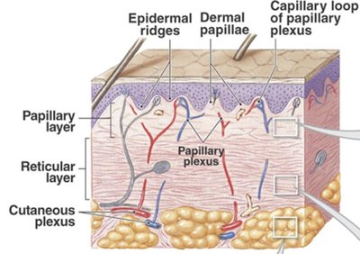
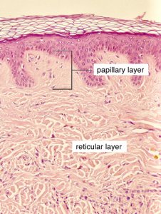
The dermis is a connective tissue layer divided into two regions:
Papillary layer: Superficial; composed of loose areolar connective tissue; contains dermal papillae that interlock with the epidermis, increasing surface area for nutrient exchange and adhesion.
Reticular layer: Deep; composed of dense irregular connective tissue; provides strength and elasticity, resists multidirectional forces.
Cells of the Dermis
Fixed cells: Fibroblasts, adipocytes, macrophages, melanocytes
Wandering cells: Mast cells, lymphocytes (immune function)
Hypodermis (Subcutaneous Layer)
The hypodermis is a layer of adipose tissue beneath the dermis. It is not technically part of the integumentary system but serves as insulation, energy storage, and shock absorption.
Accessory Structures
Hair
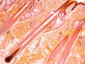
Hair is a nonliving, keratinized structure found only in thin skin. Hair follicles extend from the hypodermis and dermis to the skin surface. Hair serves protective and sensory functions.
Arrector pili muscle: Smooth muscle attached to hair follicles; contraction causes hair to stand (goosebumps).
Exocrine Glands
The skin contains several types of exocrine glands, which secrete substances onto the skin surface or into hair follicles.
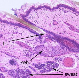
Sebaceous (oil) glands: Secrete sebum via holocrine secretion; lubricates and waterproofs skin and hair; antimicrobial properties.
Sweat (sudoriferous) glands: Two main types:
Eccrine (merocrine) glands: Produce watery, salty sweat; widely distributed; important for thermoregulation.
Apocrine glands: Found in axillae, groin, nipples; produce thicker, musky secretions; active after puberty; associated with hair follicles.
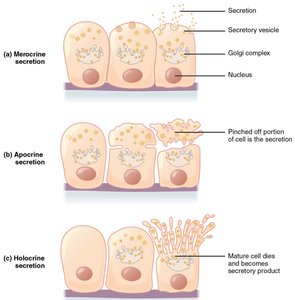
Modes of Secretion
Merocrine secretion: Product released by exocytosis (e.g., sweat glands).
Apocrine secretion: Part of the cell is pinched off (e.g., mammary glands).
Holocrine secretion: Entire cell disintegrates to release product (e.g., sebaceous glands).
Innervation and Sensory Structures
Sensory Receptors in the Skin
Merkel cells: Detect light touch; located in the stratum basale.
Tactile (Meissner's) corpuscles: Detect light touch; found in dermal papillae, especially in sensitive areas (fingers, lips).
Lamellated (Pacinian) corpuscles: Detect deep pressure and vibration; found in the reticular layer of the dermis.
Summary Table: Integumentary System Components
Component | Main Features |
|---|---|
Epidermis | Protection, water resistance, pigment production, immune defense |
Dermis | Support, elasticity, houses blood vessels and nerves |
Hair follicles | Produce hair, sensory input, protection |
Exocrine glands | Sweat production, lubrication, antimicrobial action |
Nails | Protect and support tips of fingers and toes |
Additional info: The epidermis is regenerated every 6 weeks, and the arrangement of dermal papillae and epidermal ridges contributes to fingerprints. The hypodermis is important for drug injection due to its vascularity and loose structure.
