 뒤로
뒤로The Integumentary System: Structure, Function, and Clinical Relevance
스터디 가이드 - 스마트 노트
 자료에 맞춘 맞춤형 노트, 핵심 정의, 예시, 맥락을 확장해 제공합니다.
The Integumentary System
Overview
The integumentary system is composed of the skin and its accessory structures, including hair, nails, and glands. As the largest organ system, it plays a critical role in protection, sensation, thermoregulation, excretion, and vitamin D synthesis.
Skin Structure
Main Components
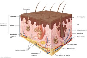
Skin (Cutaneous Membrane): Composed of two main layers:
Epidermis: Superficial, avascular layer of keratinized stratified squamous epithelium resting on a basement membrane.
Dermis: Deep, vascularized layer of loose and dense irregular connective tissue.
Accessory Structures: Hair, nails, sweat glands, sebaceous glands, sensory neurons, and arrector pili muscles.
Hypodermis (Subcutaneous Tissue): Not part of the skin; consists of loose connective and adipose tissue, highly vascularized.
Functions of the Integumentary System
Protection
Mechanical Trauma: Keratinized epithelium provides a durable, flexible barrier against stretching, pressure, and abrasion.
Pathogen Defense: Continuous skin structure, immune cells, and antimicrobial secretions (acid mantle) deter pathogen growth.
Environmental Protection: Absorbs UV radiation, secretes lipids to repel water and salts, preventing dehydration or overhydration.
Sensation
Contains sensory receptors that detect touch, temperature, pain, and other stimuli, crucial for homeostasis.
Thermoregulation
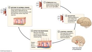
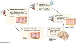
Maintains stable internal temperature via negative feedback loops.
When body temperature rises: Thermoreceptors signal the hypothalamus, which triggers sweating and vasodilation to release heat.
When body temperature falls: Thermoreceptors signal the hypothalamus, which triggers vasoconstriction to conserve heat.
Excretion
Eliminates small amounts of metabolic waste (e.g., lactic acid, urea) in sweat.
Vitamin D Synthesis
UV exposure converts a precursor in the epidermis to cholecalciferol, which is modified by the liver and kidneys to form calcitriol (active vitamin D), essential for calcium absorption.
The Epidermis
Cell Types
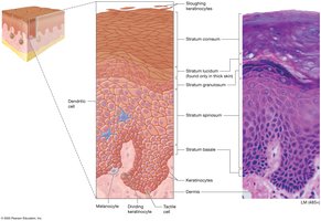
Keratinocytes: 95% of epidermal cells; produce keratin for strength and water resistance; linked by desmosomes.
Dendritic (Langerhans) Cells: Immune phagocytes in the stratum spinosum.
Tactile (Merkel) Cells: Sensory receptors for light touch in the stratum basale.
Melanocytes: Produce melanin pigment in the stratum basale.
Layers (Strata) of the Epidermis
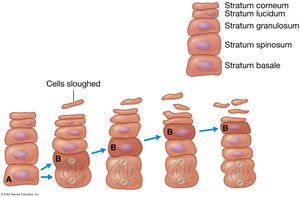
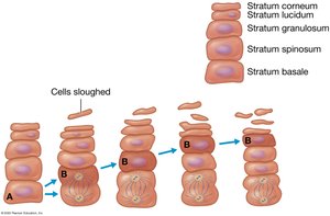
Stratum Basale (Germinativum): Deepest, single layer of stem cells; mitotically active; produces keratin and vitamin D precursors.
Stratum Spinosum: Thickest layer; spiky cells; active in keratin and vitamin D production.
Stratum Granulosum: 3–5 rows; contains keratin and membrane-coating granules; forms water barrier.
Stratum Lucidum: Clear, dead keratinocytes; only in thick skin (palms, soles).
Stratum Corneum: Multiple layers of dead, flattened keratinocytes; cells are shed (sloughed) daily.
Mnemonic for Epidermal Layers
Brilliant – Stratum Basale
Studying – Stratum Spinosum
Gives – Stratum Granulosum
Loads of – Stratum Lucidum
Confidence – Stratum Corneum
Keratinocyte Life Cycle
Cells divide in the stratum basale/spinosum, are pushed upward, and are eventually shed from the stratum corneum (cycle lasts 40–50 days).
Thick vs. Thin Skin
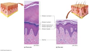
Thick Skin: All five strata; found on palms, soles; lacks hair follicles; many sweat glands.
Thin Skin: Lacks stratum lucidum; thinner stratum corneum; contains hair follicles, sweat, and sebaceous glands.
Callus: Localized thickening of stratum corneum due to repeated pressure.
The Dermis
Layers of the Dermis
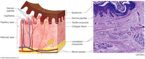
Papillary Layer: Superficial 20%; loose connective tissue; contains dermal papillae (blood supply, tactile corpuscles).
Reticular Layer: Deep 80%; dense irregular connective tissue; contains blood vessels, glands, hair follicles, lamellated corpuscles (deep pressure/vibration).
Skin Markings
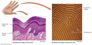
Dermal Ridges: Prominent in thick skin; form epidermal ridges (fingerprints).
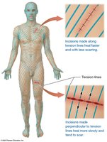
Tension (Cleavage) Lines: Gaps between collagen bundles; important for surgical incisions.
Flexure Lines: Deep creases in areas like palms.
Pigmentation and Skin Color
Melanin
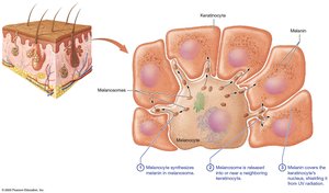
Produced by melanocytes in the stratum basale; stored in melanosomes.
Protects DNA from UV radiation; must be continually replaced.
UV exposure increases melanin synthesis (immediate darkening and delayed production).
Regulates vitamin D synthesis to maintain homeostasis.
Other Pigments
Carotene: Yellow-orange pigment from diet; accumulates in stratum corneum.
Hemoglobin: Red pigment in blood; gives skin a pinkish hue in fair-skinned individuals.
Skin Color as a Diagnostic Tool
Erythema: Increased blood flow; red appearance.
Pallor: Decreased blood flow; pale appearance.
Cyanosis: Low oxygen; bluish color.
Jaundice: High bilirubin; yellow-orange color.
Accessory Structures
Hair
Functions in protection (e.g., UV, foreign objects) and sensation.
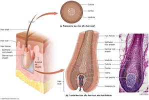
Structure: Shaft (dead cells), root (living cells), follicle, arrector pili muscle, bulb, and papilla (blood supply).
Growth: Alternates between growth and resting stages; alopecia is hair loss.
Pigment: Determined by melanin; changes with age.
Nails
Protect fingertips; tools for manipulation.
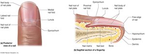
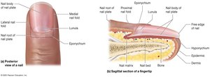
Structure: Nail plate (body and root), nail bed, matrix, folds, cuticle, lunula.
Growth: Continuous; lunula is the opaque region with more keratin.
Glands
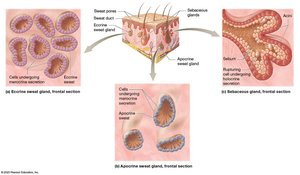
Eccrine Sweat Glands: Most common; thermoregulation; watery secretion via pores.
Apocrine Sweat Glands: Axillae, anal, areolae; thick, protein-rich secretion onto hair follicles; odor develops after bacterial metabolism.
Ceruminous Glands: Modified apocrine glands; produce ear wax (cerumen).
Mammary Glands: Modified sweat glands; produce milk.
Sebaceous Glands: Secrete sebum (oily, hydrophobic barrier) via holocrine secretion; increases at puberty.
Clinical Aspects
Burns
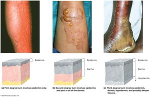
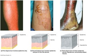
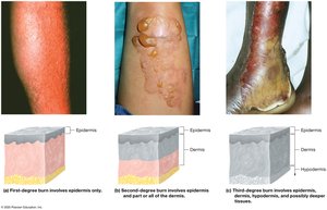
First-Degree: Only epidermis; erythema, minor pain, no blisters.
Second-Degree: Epidermis and part/all of dermis; pain, blisters, possible scarring.
Third-Degree: Full thickness; all skin layers and possibly deeper; not initially painful due to nerve damage; risk of dehydration and infection; may require grafting.
Rule of Nines
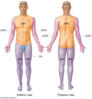
Used to estimate burn surface area for fluid replacement; body divided into regions representing 9% each (genital area is 1%).
Skin Cancer
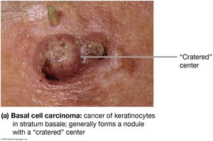
Basal Cell Carcinoma: Most common; arises from stratum basale; rarely metastasizes.
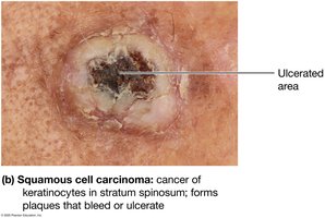
Squamous Cell Carcinoma: From stratum spinosum; may metastasize; forms scaly plaques.
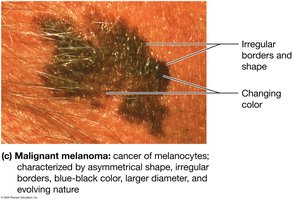
Malignant Melanoma: Most dangerous; from melanocytes; high metastatic potential; identified by ABCDE rule (Asymmetry, Border, Color, Diameter, Evolving).
Summary Table: Epidermal Layers
Layer | Location | Key Features |
|---|---|---|
Stratum Basale | Deepest | Mitotically active stem cells, melanocytes, tactile cells |
Stratum Spinosum | Above basale | Thickest, keratin production, dendritic cells |
Stratum Granulosum | Middle | Granules, water barrier formation |
Stratum Lucidum | Thick skin only | Clear, dead keratinocytes |
Stratum Corneum | Superficial | Dead, flattened keratinocytes, sloughing |
Additional info:
Cellulite is a normal anatomical variation, not a disease.
Topical medications can be absorbed through the skin if they are nonpolar; this is also a route for some toxins.
Wrinkles result from decreased collagen, elastic fibers, and adipose tissue; accelerated by UV exposure and smoking.
Acne is caused by sebum and dead cell accumulation in sebaceous glands, often influenced by hormones.
