 뒤로
뒤로The Integumentary System: Structure, Function, and Clinical Relevance
스터디 가이드 - 스마트 노트
 자료에 맞춘 맞춤형 노트, 핵심 정의, 예시, 맥락을 확장해 제공합니다.
The Integumentary System
Overview and Components
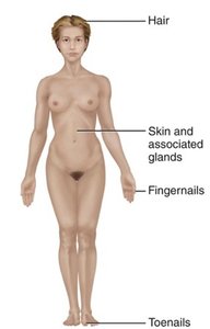
The integumentary system is the body's primary protective barrier, consisting of the skin, hair, nails, sweat glands, sebaceous (oil) glands, and sensory receptors. It plays a crucial role in maintaining homeostasis, protecting internal organs, and regulating body temperature.
Skin: The largest organ, covering the entire body.
Hair: Provides protection and sensory input.
Nails: Protects the distal phalanges and aids in grasping.
Sweat glands: Regulate temperature and excrete wastes.
Sebaceous glands: Lubricate skin and hair.
Functions of the Integumentary System
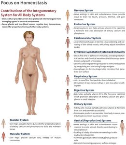
The integumentary system serves several essential functions:
Protection: Guards against microorganisms, abrasions, temperature extremes, and harmful chemicals.
Temperature Regulation: Maintains stable internal body temperature through sweat and blood flow.
Sensory Reception: Contains receptors for touch, pain, temperature, and pressure.
Metabolic Functions: Synthesizes vitamin D and aids in hormone activation.
Excretion: Removes waste products via sweat.
Structure of the Skin
Layers of the Skin
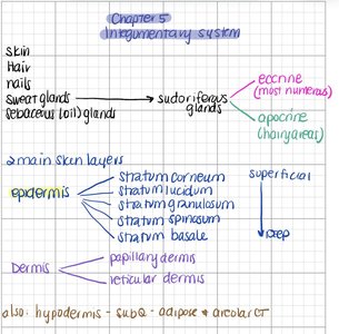
The skin is composed of three main layers:
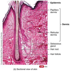
Epidermis: The most superficial layer, made of keratinized stratified squamous epithelium.
Dermis: Deep to the epidermis, composed of connective tissue with collagen and elastic fibers.
Hypodermis (Subcutaneous Layer): Not technically part of the skin; consists of areolar and adipose tissue.
Epidermis: Cell Types and Layers
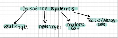
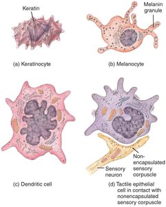
The epidermis contains four principal cell types:
Keratinocytes: Produce keratin, providing strength and waterproofing.
Melanocytes: Produce melanin, protecting against UV radiation.
Dendritic (Langerhans) cells: Immune cells that patrol the epidermis.
Tactile (Merkel) cells: Sensory receptors for touch.
Layers of the Epidermis (from deep to superficial):
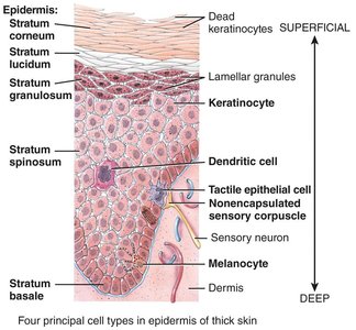
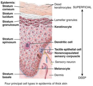
Stratum basale: Single row of stem cells; site of cell division and melanocytes.
Stratum spinosum: Several layers of keratinocytes; contains dendritic cells and melanosomes.
Stratum granulosum: Cells flatten and keratinization begins; lamellar granules provide water resistance.
Stratum lucidum: Present only in thick skin; clear, flat, dead keratinocytes.
Stratum corneum: 20–30 rows of dead, keratinized cells; provides protection.
Dermis: Structure and Layers
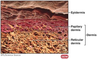
The dermis is a strong, flexible connective tissue layer containing nerves, blood vessels, lymphatic vessels, hair follicles, and glands. It is divided into two layers:
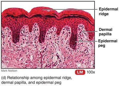
Papillary layer: Superficial, areolar connective tissue; contains dermal papillae, capillary loops, and tactile corpuscles.
Reticular layer: Deep, dense irregular connective tissue; contains collagen and elastic fibers, adipose cells, and skin appendages.
Special Features: Epidermal Ridges and Flexure Lines
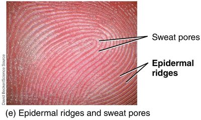
Epidermal ridges: Enhance gripping ability and contribute to fingerprints; formed by dermal papillae lying atop dermal ridges in thick skin.
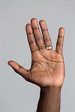
Flexure lines: Dermal folds at or near joints, visible on hands, wrists, fingers, soles, and toes.
Pigmentation and Skin Tone
Skin Pigments
Three pigments contribute to skin tone:
Melanin: Produced by melanocytes; shields DNA from UV damage.
Hemoglobin: Red pigment in blood; gives pinkish hue to fair skin.
Carotene: Yellow-orange pigment stored in the stratum corneum and adipose tissue; can be converted to vitamin A.
Accessory Structures
Hair
Hair consists of dead, keratinized cells and is present on most body surfaces except palms, soles, lips, nipples, and parts of external genitalia.
Functions: Protection, sensory input, regulation of heat loss.
Structure: Shaft (above skin), root (penetrates dermis), follicle, epithelial root sheath, dermal root sheath.
Hair shaft components: Medulla, cortex, cuticle.
Pigmentation: Determined by melanocyte activity and melanin type.
Nails
Nails are modifications of the epidermis containing hard keratin.
Functions: Protect distal phalanges, aid in grasping.
Structure: Free edge, nail plate, root, nail bed, nail matrix, nail folds, eponychium (cuticle), hyponychium, lunule.
Glands
The skin contains three main types of glands:
Sebaceous (oil) glands: Connected to hair follicles; secrete sebum.
Sudoriferous (sweat) glands: Eccrine (most numerous, thermoregulation) and apocrine (hairy areas, body odor).
Ceruminous glands: Modified sweat glands in the ear canal; produce cerumen (earwax).
Skin as a Barrier
Types of Barriers
Chemical barrier: Sweat, sebum, acid mantle, melanin.
Physical barrier: Keratinized cells, glycolipids, limited penetration by chemicals.
Biological barrier: Dendritic cells, macrophages, DNA absorbs UV radiation.
Thermoregulation and Sensation
Thermoregulation
Insensible perspiration: Unnoticeable sweat under normal conditions.
Sensible perspiration: Noticeable sweat during elevated body temperature.
Cold response: Dermal blood vessels constrict to conserve heat.
Cutaneous Sensations
Tactile sensations: Touch, pressure, vibration, tickle.
Thermal sensations: Warm, cool.
Pain: Nociceptors respond to damaging stimuli.
Wound Healing and Clinical Relevance
Wound Healing
Epidermal wound healing: Superficial wounds affect only the epidermis.
Deep wound healing: Involves inflammatory, migratory, proliferative, and maturation phases.
Skin Cancer
Basal cell carcinoma: Most common, least dangerous.
Squamous cell carcinoma: Second most common.
Malignant melanoma: Most dangerous; early warning signs include asymmetry, irregular border, uneven color, diameter >6mm, evolving shape.
Burns
First-degree: Epidermal damage only.
Second-degree: Epidermal and upper dermal damage; blisters.
Third-degree: Entire thickness of skin; requires skin grafting.
Development and Age-Associated Changes
Development
Epidermis: Develops from ectoderm.
Dermis: Develops from mesoderm.
Accessory structures: Nails, hair, and glands are epidermal derivatives.
Age-Associated Changes
Wrinkles, dehydration, decreased sweat production, loss of melanocytes (gray hair, atypical pigmentation), loss of subcutaneous fat, decreased skin thickness, brittle nails.
Clinical Connections
Pressure Ulcers
Increased susceptibility with age due to decreased blood flow and shedding of epithelium.
Albinism and Vitiligo
Albinism: Inherited inability to produce melanin.
Vitiligo: Loss of melanocytes from patches of skin due to immune malfunction.
Comparative Tables
Gland Comparison
Feature | Sebaceous Glands | Eccrine Sweat Glands | Apocrine Sweat Glands | Ceruminous Glands |
|---|---|---|---|---|
Distribution | Lips, glans penis, labia minora, tarsal glands; small in trunk/limbs; absent in palms/soles | Throughout most skin, especially forehead, palms, soles | Axillae, groin, areolae, bearded face, clitoris, labia minora | External acoustic meatus |
Location of secretory portion | Dermis | Mostly reticular dermis | Reticular dermis/superficial subcutaneous tissue | Subcutaneous tissue |
Termination of excretory duct | Hair follicle | Surface of epidermis | Hair follicles | Surface of external auditory canal or ducts of sebaceous glands |
Secretion | Sebum (triglycerides, cholesterol, proteins, inorganic salts) | Perspiration (water, ions, urea, uric acid, ammonia, amino acids, glucose, lactic acid) | Perspiration plus lipids and proteins | Cerumen (waxy material) |
Functions | Prevent hair drying, water loss, keep skin soft, inhibit bacteria | Regulate temperature, waste removal, emotional stress | Emotional stress, sexual excitement | Impede foreign bodies, waterproof canal, prevent microbes |
Onset of function | Puberty | Birth | Puberty | Birth |
Thin vs. Thick Skin
Feature | Thin Skin | Thick Skin |
|---|---|---|
Distribution | All except palms, palmar surfaces of digits, soles, plantar surfaces of toes | Palms, palmar surfaces of digits, soles, plantar surfaces of toes |
Epidermal thickness | 0.10–0.15 mm | 0.6–4.5 mm (thicker stratum corneum) |
Epidermal strata | Stratum lucidum lacking; thinner spinosum and corneum | Stratum lucidum present; thicker spinosum and corneum |
Epidermal ridges | Poorly developed, fewer, less organized dermal papillae | Well developed, numerous dermal papillae in parallel rows |
Hair follicles/arrector muscles | Present | Absent |
Sebaceous glands | Present | Absent |
Sudoriferous glands | Fewer | More numerous |
Sensory receptors | Sparser | Denser |
