뒤로
뒤로The Integumentary System: Structure, Function, and Clinical Relevance
스터디 가이드 - 스마트 노트
 자료에 맞춘 맞춤형 노트, 핵심 정의, 예시, 맥락을 확장해 제공합니다.
The Integumentary System
Overview and Functions
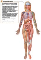
The integumentary system is the body's largest organ system, consisting of the skin and its accessory structures. It serves as the primary barrier between the internal environment and the external world, providing protection, sensory input, and regulatory functions.
Physical protection: Shields underlying tissues from mechanical damage, pathogens, and chemical exposure.
Thermoregulation: Regulates body temperature through sweat production and blood flow.
Sensory reception: Contains receptors for touch, pressure, pain, and temperature.
Excretion: Removes metabolic wastes through sweat.
Synthesis of vitamin D: Initiates vitamin D production when exposed to sunlight.
Storage of lipids: Stores energy in the form of fat in the subcutaneous layer.
Components of the Integumentary System
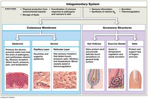
Cutaneous Membrane
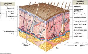
Epidermis: The outermost, avascular layer composed of stratified squamous epithelium.
Dermis: The deeper, vascular layer containing connective tissue, blood vessels, and nerves.
Accessory Structures
Hair follicles
Exocrine glands (sebaceous and sweat glands)
Nails
Epidermis
Structure and Cell Types
The epidermis is the outermost layer of the skin, composed primarily of keratinocytes. It is avascular, receiving nutrients and oxygen by diffusion from the underlying dermis.
Keratinocytes: Main cell type, produce keratin (a tough, fibrous protein).
Stratified squamous epithelium: Provides durability and protection.
Layers of the Epidermis
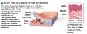
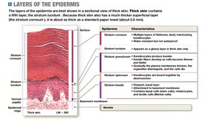
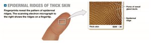
The epidermis consists of several distinct layers, each with specialized functions. In thick skin (palms, soles), all five layers are present; in thin skin, the stratum lucidum is absent.
Layer | Characteristics |
|---|---|
Stratum basale | Deepest layer; contains stem cells (basal cells), melanocytes, and tactile (Merkel) cells |
Stratum spinosum | Several layers of keratinocytes; some cell division; contains dendritic (Langerhans) cells |
Stratum granulosum | Keratinocytes stop dividing, begin keratinization |
Stratum lucidum | Present only in thick skin; clear layer of dead keratinocytes |
Stratum corneum | Multiple layers of dead, keratinized cells; forms the exposed surface |
Specialized Cells of the Stratum Basale
Basal cells: Stem cells that divide to produce new keratinocytes.
Tactile (Merkel) cells: Sensory receptors for touch.
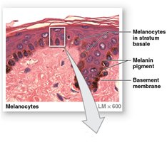
Melanocytes: Synthesize the pigment melanin, which protects against UV radiation.
Dermis
Structure and Layers
The dermis is the connective tissue layer beneath the epidermis, providing structural support and housing blood vessels, nerves, and accessory structures.
Papillary layer: Superficial, composed of areolar tissue; contains capillaries, lymphatics, and sensory neurons.
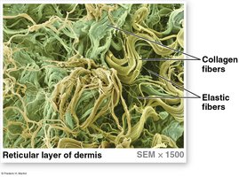
Reticular layer: Deeper, composed of dense irregular connective tissue with collagen and elastic fibers.
Sensory Receptors in the Dermis
Tactile (Meissner) corpuscles: Detect light touch, located in dermal papillae.
Lamellar (Pacinian) corpuscles: Detect deep pressure and vibration, located in the reticular layer.
Subcutaneous Layer (Hypodermis)
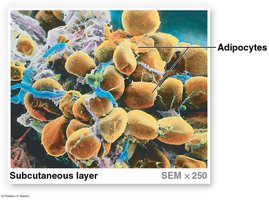
The subcutaneous layer is not technically part of the skin but connects the dermis to underlying tissues. It consists mainly of adipose tissue, providing insulation, energy storage, and cushioning.
Adipocytes: Fat cells that store energy.
Clinical relevance: Common site for subcutaneous injections.
Skin Color
Melanin and Its Role
Skin color is primarily determined by the amount and type of melanin produced by melanocytes in the stratum basale. Melanin is stored in melanosomes and transferred to keratinocytes.
Pheomelanin: Red-yellow pigment.
Eumelanin: Brown-black pigment.
Function: Melanin protects DNA from ultraviolet (UV) radiation damage.
Clinical Note: Skin Disorders
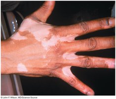
Vitiligo: Loss of melanocytes, resulting in patchy loss of skin color.
Skin cancer: Includes basal cell carcinoma (most common, originates in stratum basale), squamous cell carcinoma (arises from squamous cells), and malignant melanoma (from melanocytes, highly metastatic).
Accessory Structures of the Skin
Hair and Hair Follicles
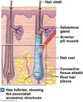
Hair is produced by hair follicles, which are invaginations of the epidermis into the dermis. Hair serves protective and sensory functions.
Hair matrix: Basal cells divide to produce hair cells, which keratinize and die as they are pushed upward.
Growth cycle: Includes active growth, regression, and shedding phases.
Exocrine Glands of the Skin
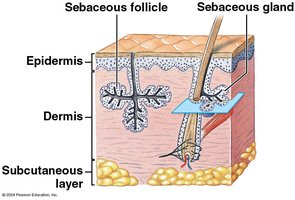
Sebaceous glands: Secrete sebum (oily substance) into hair follicles for lubrication and waterproofing.
Sweat glands: Two main types:
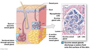
Eccrine (merocrine) glands: Secrete watery sweat directly onto skin surface; important for thermoregulation.
Apocrine glands: Found in armpits and pubic region; secrete a thicker, odor-producing fluid beginning at puberty.
Nails
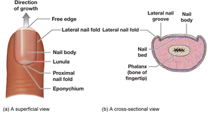
Nails are protective structures composed of dead, keratinized epidermal cells. They protect the tips of fingers and toes and aid in manipulation of objects.
Nail body: Visible part of the nail.
Nail bed: Skin beneath the nail body.
Lunula: Crescent-shaped area at the base of the nail.
Repair of Injury to the Integument
Phases of Skin Repair
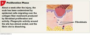
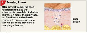
Skin repair after injury involves a series of coordinated phases:
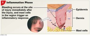
Inflammation phase: Bleeding and inflammatory response initiated by mast cells.
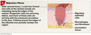
Migration phase: Formation of a scab; migration of epithelial cells and arrival of phagocytes and fibroblasts.
Proliferation phase: Epidermal cells proliferate; fibroblasts produce collagen fibers; phagocytosis decreases.
Scarring phase: Scab is shed; fibroblasts continue to strengthen the area, forming scar tissue.
Integration with Other Body Systems
The integumentary system interacts with other organ systems to maintain homeostasis. For example, it works with the nervous system for sensory input, the circulatory system for thermoregulation, and the immune system for defense against pathogens.