 뒤로
뒤로The Integumentary System: Structure, Function, and Clinical Aspects
스터디 가이드 - 스마트 노트
 자료에 맞춘 맞춤형 노트, 핵심 정의, 예시, 맥락을 확장해 제공합니다.
The Integumentary System
Overview and Functions
The integumentary system is the body's largest organ system, primarily composed of the skin and its associated structures. It serves as a protective barrier, regulates temperature, and provides sensory information.
Protection: Shields internal tissues from environmental hazards, pathogens, and physical trauma.
Thermoregulation: Controls body temperature through sweat production and blood vessel dilation/constriction.
Sensory Reception: Detects touch, pressure, pain, and temperature via specialized receptors.
Excretion: Removes waste products through sweat.
Vitamin D Synthesis: Initiates synthesis of vitamin D3 when exposed to sunlight.
Components of the Integumentary System
The system consists of the cutaneous membrane (skin), accessory structures (hair, nails, glands), and the subcutaneous layer.
Skin: Composed of the epidermis and dermis.
Accessory Structures: Includes hair follicles, exocrine glands, and nails.
Subcutaneous Layer (Hypodermis): Attaches skin to deeper tissues and stores lipids.
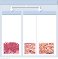
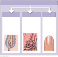
Structure of the Skin
Layers of the Skin
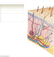
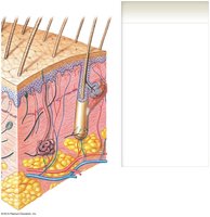
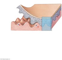
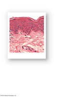
The skin is organized into three main layers:
Epidermis: The outermost layer, composed of stratified squamous epithelium.
Dermis: Beneath the epidermis, contains connective tissue, blood vessels, nerves, and accessory structures.
Subcutaneous Layer (Hypodermis): Deepest layer, primarily adipose tissue for insulation and energy storage.
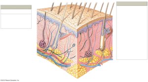
Dermis: Papillary and Reticular Layers
The dermis is divided into two layers:
Papillary Layer: Loose connective tissue, contains capillaries and sensory neurons.
Reticular Layer: Dense, irregular connective tissue, provides strength and elasticity.
Epidermis: Structure and Cell Types
Layers of the Epidermis
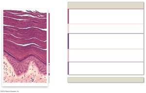
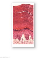
The epidermis consists of several distinct layers, especially prominent in thick skin:
Stratum Corneum: Multiple layers of dead, keratinized cells; provides water resistance.
Stratum Lucidum: Found only in thick skin (palms, soles); clear, glassy layer.
Stratum Granulosum: Keratinocytes produce keratin and keratohyalin; cells begin to die.
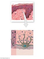
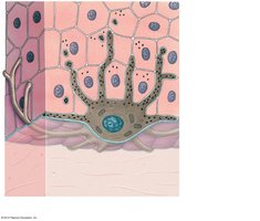
Stratum Spinosum: Keratinocytes bound together; contains Langerhans cells and melanocytes.
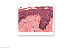
Stratum Basale: Deepest layer; contains stem cells, melanocytes, and Merkel cells.
Cell Types in the Epidermis
Keratinocytes: Produce keratin, a tough, fibrous protein.
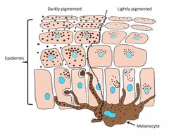
Melanocytes: Produce melanin, responsible for skin color and UV protection.
Merkel Cells: Sensory cells for touch.
Langerhans Cells: Immune cells, protect against pathogens and cancer.
Skin Types: Thin vs. Thick Skin
Differences Between Thin and Thick Skin
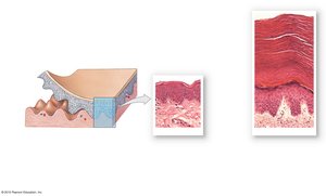
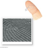
Thick Skin: Found on palms and soles; contains all five epidermal layers, including stratum lucidum.
Thin Skin: Covers most of the body; lacks stratum lucidum, thinner stratum corneum.
Accessory Structures
Hair and Hair Follicles
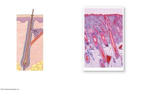
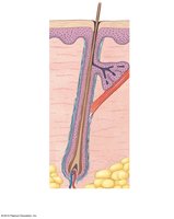
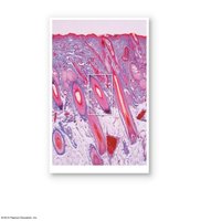
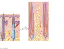
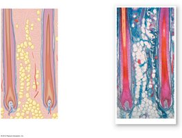
Hair is produced by follicles located in the dermis and serves multiple functions.
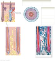
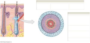
Structure: Composed of medulla (soft keratin), cortex (hard keratin), and cuticle (hard keratin).
Follicle: Contains internal and external root sheaths, glassy membrane, and connective tissue sheath.
Functions: Protection, insulation, sensory input, and thermoregulation (arrector pili muscle causes goosebumps).
Nails
Nails are protective structures at the distal tips of fingers and toes.
Structure: Composed of nail body, free edge, lunula, nail root, and cuticle (eponychium).
Function: Protects and stiffens distal tips, aids in manipulation and sensation.
Exocrine Glands
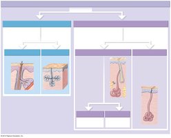
The skin contains several types of exocrine glands:
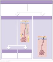
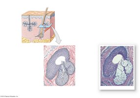
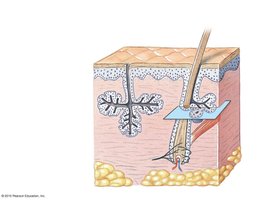
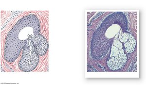
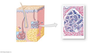
Sebaceous Glands: Secrete sebum (oily lipid) for lubrication and antibacterial action; associated with hair follicles or open directly onto skin.
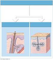
Sweat Glands:
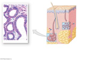
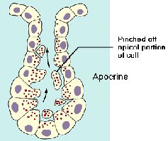
Apocrine: Limited distribution (axillae, groin, nipples); produce viscous secretion, possible role in communication.
Merocrine (Eccrine): Widespread; produce watery sweat for thermoregulation and excretion.
Ceruminous Glands: Produce waxy cerumen in external ear canal.
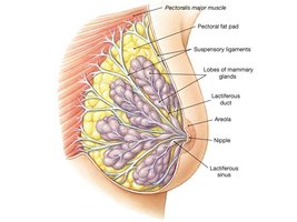
Mammary Glands: Specialized apocrine glands for milk production.
Skin Pigmentation
Melanin, Hemoglobin, and Carotene
Skin color is determined by the presence of melanin, hemoglobin, and carotene.
Melanin: Produced by melanocytes; protects against UV radiation.
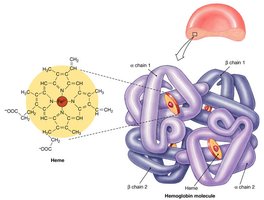
Hemoglobin: Oxygenated blood imparts a reddish hue.
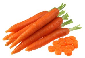
Carotene: Yellow-orange pigment from diet, accumulates in epidermal cells.

Clinical Aspects
Repairing Injuries to the Skin
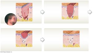
Skin injury initiates a complex repair process involving inflammation, cell migration, and tissue remodeling.
Inflammatory Response: Bleeding and mast cell activation.
Scab Formation: Migration of epithelial cells and phagocytic activity.
Tissue Repair: Fibroblasts produce granulation tissue and scar formation.
Remodeling: Scar tissue elevates epidermis; healing continues over weeks.
Aging and the Skin
Aging causes structural and functional changes in the skin:
Thinner Epidermis and Dermis: Increased fragility and tendency to wrinkle.
Reduced Melanocyte Activity: Pale skin, decreased UV protection.
Decreased Gland Activity: Dry skin, reduced thermoregulation.
Impaired Healing: Slower repair and increased risk of infection.
Summary Table: Layers and Structures of the Skin
Layer | Main Tissue | Key Structures |
|---|---|---|
Epidermis | Stratified squamous epithelium | Keratinocytes, melanocytes, Merkel cells, Langerhans cells |
Dermis (Papillary) | Loose connective tissue | Capillaries, sensory neurons, dermal papillae |
Dermis (Reticular) | Dense irregular connective tissue | Collagen fibers, elastic fibers, glands, hair follicles |
Hypodermis | Adipose tissue | Fat cells, blood vessels |
Summary Table: Accessory Structures
Structure | Function |
|---|---|
Hair | Protection, sensory input, thermoregulation |
Nails | Protect and stiffen distal tips of digits |
Sebaceous Glands | Lubricate skin and hair, antibacterial action |
Sweat Glands | Thermoregulation, excretion |
Key Equations
Water Loss by Insensible Perspiration
Hair Growth Rate
Additional info:
Some histological images and diagrams were inferred to represent the main skin layers and accessory structures based on context.
Tables were reconstructed to summarize the main tissue types and functions of skin layers and accessory structures.
