뒤로
뒤로The Integumentary System: Structure, Function, and Clinical Relevance
스터디 가이드 - 스마트 노트
 자료에 맞춘 맞춤형 노트, 핵심 정의, 예시, 맥락을 확장해 제공합니다.
The Integumentary System
Overview
The integumentary system is composed of the skin and its accessory structures, including hair, nails, and glands. As the largest organ system, it plays a critical role in protection, sensation, thermoregulation, excretion, and vitamin D synthesis.
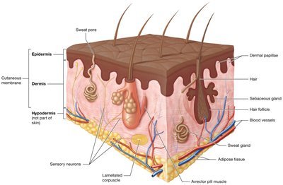
Skin (Cutaneous Membrane): Comprises 10-15% of total body weight and consists of two main layers: the superficial epidermis and the deeper dermis.
Accessory Structures: Include hair, nails, sweat glands, and sebaceous glands.
Hypodermis (Subcutaneous Tissue): Lies below the dermis, composed of loose connective and adipose tissue, and is highly vascularized but not considered part of the skin.
Functions of the Integumentary System
Protection
Mechanical Trauma: Keratinized stratified squamous epithelium provides a durable, flexible barrier against stretching, pressure, and abrasion.
Pathogen Defense: The skin's continuous structure, immune cells, and antimicrobial secretions (acid mantle) deter pathogen invasion.
Environmental Protection: Absorbs UV radiation and prevents water loss/gain via lipid-based secretions.
Sensation
Contains sensory receptors that detect touch, temperature, pain, and other stimuli, crucial for homeostasis.
Thermoregulation
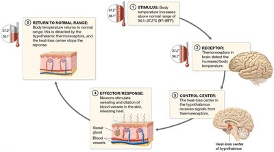
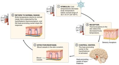
Maintains stable internal temperature through negative feedback loops:
When body temperature rises: Thermoreceptors signal the hypothalamus, which stimulates sweating and vasodilation to release heat.
When body temperature falls: Thermoreceptors signal the hypothalamus, which stimulates vasoconstriction to conserve heat.
Excretion
Eliminates small amounts of metabolic waste (e.g., lactic acid, urea) in sweat.
Vitamin D Synthesis
UV exposure converts a precursor in the epidermis to cholecalciferol (vitamin D3), which is modified by the liver and kidneys to form calcitriol, essential for calcium absorption.
Skin Structure
The Epidermis
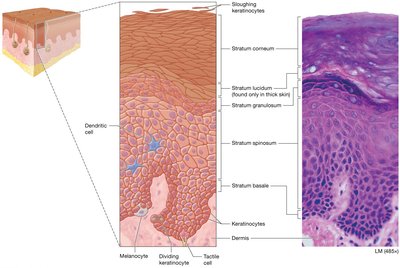
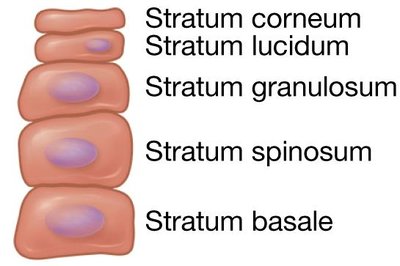
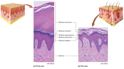
The epidermis is the superficial layer of the skin, composed primarily of keratinocytes arranged in up to five distinct layers (strata):
Stratum Basale (Germinativum): Deepest layer; single row of stem cells; mitotically active; produces keratin and vitamin D precursors.
Stratum Spinosum: Thickest layer; spiny-shaped cells; active in keratin and vitamin D production.
Stratum Granulosum: 3-5 rows of cells with granules; forms water barrier.
Stratum Lucidum: Clear layer of dead keratinocytes; present only in thick skin.
Stratum Corneum: Multiple layers of dead, flattened keratinocytes; provides durable, protective surface.
Keratinocyte Life Cycle
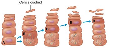
Keratinocytes originate in the stratum basale and are pushed upward, differentiating as they move through the strata, and are eventually sloughed off in the stratum corneum (process takes 40-50 days).
Other Epidermal Cells
Dendritic (Langerhans) Cells: Immune phagocytes in the stratum spinosum.
Tactile (Merkel) Cells: Sensory receptors for light touch in the stratum basale.
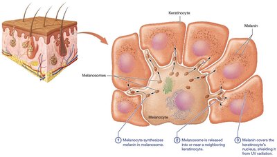
Melanocytes: Produce melanin pigment in the stratum basale.
Thick vs. Thin Skin
Thick Skin: All five strata; found on palms, soles, and palmar/plantar surfaces; lacks hair follicles but has many sweat glands.
Thin Skin: Lacks stratum lucidum; thinner stratum corneum; contains hair follicles, sweat, and sebaceous glands.
Callus: Localized thickening of the stratum corneum due to repeated pressure.
The Dermis
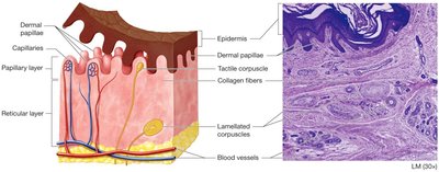
The dermis is the deep layer of the skin, providing structural support and housing blood vessels, nerves, and accessory structures. It is divided into two layers:
Papillary Layer: Superficial; loose connective tissue; contains dermal papillae with capillaries and tactile (Meissner) corpuscles for light touch.
Reticular Layer: Deep; dense irregular connective tissue; contains blood vessels, sweat and sebaceous glands, hair follicles, and lamellated (Pacinian) corpuscles for deep pressure and vibration.
Skin Markings
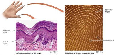
Dermal Ridges: Prominent in thick skin; form epidermal ridges (fingerprints) unique to each individual.
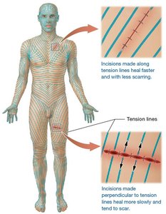
Tension (Cleavage) Lines: Gaps between collagen bundles; important for surgical incisions to minimize scarring.
Flexure Lines: Deep creases in areas such as the palms.
Skin Wrinkles
Result from decreased collagen, elastic fibers, proteoglycans, and adipose tissue with age; accelerated by UV exposure and smoking.
Temporary treatments include Botox, fillers, peels, and topical creams (with limited efficacy).


Pigmentation and Skin Color
Melanin
Produced by melanocytes in the stratum basale; packaged in melanosomes and transferred to keratinocytes.
Shields DNA from UV radiation; synthesis increases with UV exposure.
Skin color differences are due to melanin amount and type, not melanocyte number.
Common variations: freckles, moles, albinism (lack of tyrosinase enzyme).
Other Pigments
Carotene: Yellow-orange pigment from diet; accumulates in stratum corneum.
Hemoglobin: Red pigment in blood; gives pinkish hue to fair skin.
Skin Color as a Diagnostic Tool
Erythema: Increased blood flow; red appearance.
Pallor: Decreased blood flow; pale appearance.
Cyanosis: Low oxygen; bluish appearance.
Jaundice: High bilirubin; yellow-orange appearance.
Accessory Structures of the Integument
Hair
Found on most body surfaces except thick skin, lips, and parts of the genitalia.
Functions: protection (e.g., UV, foreign objects), sensation (via sensory neurons).
Structure:
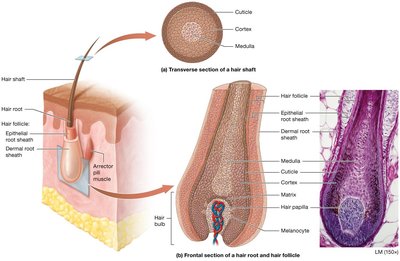
Hair Shaft: Dead keratinized cells; three regions—medulla (soft keratin), cortex (hard keratin), cuticle (hard keratin).
Hair Root: Living cells; embedded in the dermis within the hair follicle; associated with arrector pili muscle (piloerection/goosebumps).
Hair Bulb: Base of the root; contains hair papilla with capillaries.
Growth: Alternates between growth and resting stages; alopecia is hair loss due to follicle death.
Pigment and Texture: Determined by melanin type and amount; changes with age.
Nails
Hard structures at the ends of digits; protect and aid in manipulation.
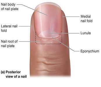
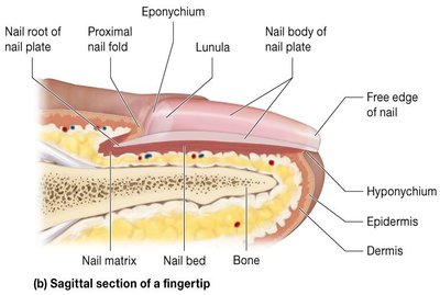
Structure: Nail plate (body and root), nail matrix (growth region), nail folds, cuticle (eponychium), hyponychium.
Growth: Continuous; fingernails grow faster than toenails.
Pigment: Nails are translucent; lunula is the whitish crescent due to thickened keratin.
Glands
Sweat (Sudoriferous) Glands:
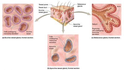
Eccrine: Most common; secrete watery sweat for thermoregulation.
Apocrine: Found in axillae, anal, and areolar regions; secrete protein-rich sweat onto hair follicles; odor develops when metabolized by bacteria.
Ceruminous: Modified apocrine glands in the ear; produce cerumen (ear wax).
Mammary: Specialized for milk production.
Sebaceous Glands: Secrete sebum (oily, waxy substance) via holocrine secretion; lubricates and waterproofs skin and hair; activity increases at puberty.
Acne
Results from accumulation of sebum and dead cells in sebaceous glands, forming comedones (blackheads); infection leads to pustules (pimples).
More common in males due to androgen influence.

Wounds, Burns, and Skin Cancer
Wounds
Any disruption in skin integrity; includes lacerations, burns, and cancers.
Burns
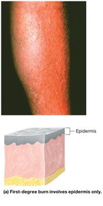
First-Degree: Only epidermis; erythema and minor pain; no permanent damage.
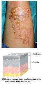
Second-Degree: Epidermis and part/all of dermis; pain, blistering, possible scarring.
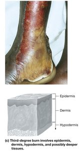
Third-Degree: Full thickness (epidermis, dermis, hypodermis, possibly deeper); not initially painful due to nerve damage; risk of dehydration and infection; may require skin grafts.
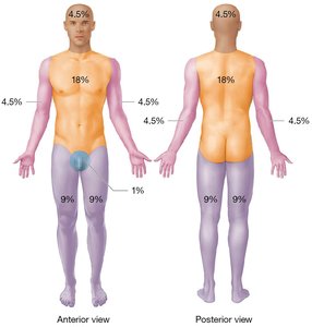
Rule of Nines
Used to estimate the percentage of body surface area affected by burns for fluid resuscitation.
Body divided into 11 areas, each representing 9% (genital area is 1%).
Skin Cancer
Results from mutations causing uncontrolled cell division (tumors); can metastasize.
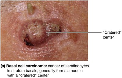
Basal Cell Carcinoma: Most common; arises from stratum basale keratinocytes; rarely metastasizes.
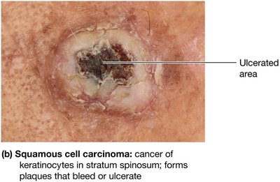
Squamous Cell Carcinoma: Second most common; arises from stratum spinosum keratinocytes; may metastasize.
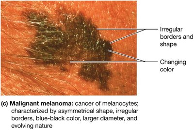
Malignant Melanoma: Least common but most dangerous; arises from melanocytes; high metastatic potential; diagnosed using the ABCDE rule (Asymmetry, Border, Color, Diameter, Evolving).
Type | Origin | Appearance | Metastasis |
|---|---|---|---|
Basal Cell Carcinoma | Stratum basale keratinocytes | Cratered nodule | Rare |
Squamous Cell Carcinoma | Stratum spinosum keratinocytes | Scaly plaque, ulceration | Possible |
Malignant Melanoma | Melanocytes | Irregular, multicolored, evolving lesion | Common |