 뒤로
뒤로The Language of Anatomy: Anatomical Position, Terminology, Planes, and Body Cavities
스터디 가이드 - 스마트 노트
 자료에 맞춘 맞춤형 노트, 핵심 정의, 예시, 맥락을 확장해 제공합니다.
The Language of Anatomy
Introduction
The study of anatomy requires a precise and universally accepted language to describe the locations and relationships of body structures. This foundational terminology allows for clear communication among healthcare professionals and students. This section introduces the anatomical position, directional terms, body planes, and body cavities.
Anatomical Position and Surface Anatomy
Anatomical Position
The anatomical position is the standard reference point in anatomy. In this position, the body is erect, feet slightly apart, head and toes pointed forward, and arms at the sides with palms facing forward. This position is essential because all directional terms refer to the body as if it were in this position, regardless of its actual posture.
Importance: Provides a consistent frame of reference for anatomical terminology.
Note: The hands are held unnaturally forward, not cupped toward the thighs.
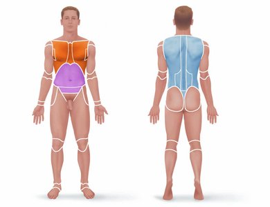
Surface Anatomy
Surface anatomy divides the body into two major regions:
Axial region: Head, neck, and trunk (the axis of the body).
Appendicular region: Limbs and their attachments to the axis.
Key anterior and posterior body landmarks include regions such as abdominal, acromial, brachial, cervical, femoral, lumbar, and many others. These terms are used to specify precise locations on the body.
Body Orientation and Direction
Directional Terms
Directional terms describe the positions of structures relative to other structures or locations in the body. These terms are always used in reference to the anatomical position.
Superior (cranial)/Inferior (caudal): Above/below along the body’s long axis.
Anterior (ventral)/Posterior (dorsal): Front/back of the body.
Medial/Lateral: Toward/away from the midline of the body.
Proximal/Distal: Nearer/farther from the trunk or point of attachment (mainly for limbs).
Superficial (external)/Deep (internal): Toward/away from the body surface.
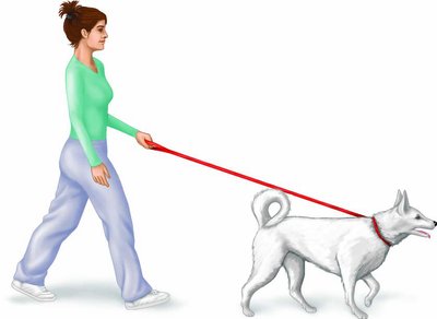
Note: In quadrupeds, some terms (e.g., dorsal/ventral) have different meanings compared to humans.
Body Planes and Sections
Major Body Planes
To study internal structures, the body is often sectioned along imaginary flat surfaces called planes. The three primary planes are:
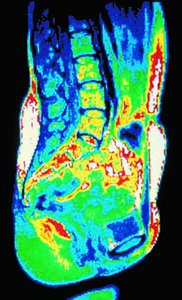
Sagittal plane: Divides the body into right and left parts. If exactly at the midline, it is the midsagittal plane.
Frontal (coronal) plane: Divides the body into anterior and posterior parts.
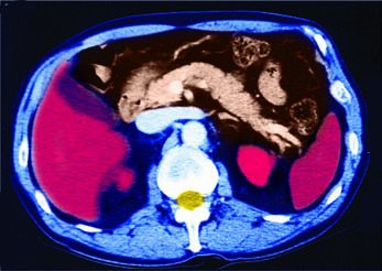
Transverse (horizontal) plane: Divides the body into superior and inferior parts; sections are called cross sections.
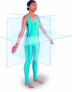
Sections through these planes provide different views of organs and structures, which is important for imaging techniques such as MRI and CT scans.
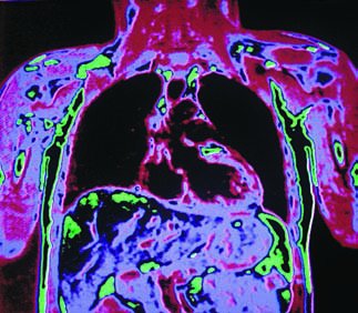
Body Cavities
Major Body Cavities
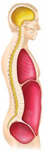
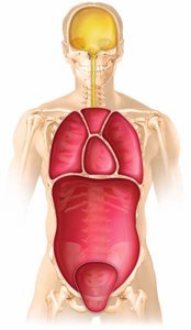
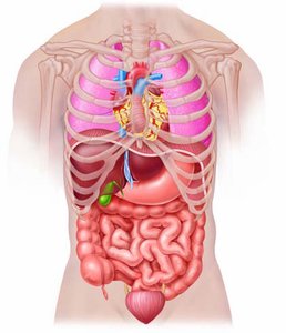
The body contains two main sets of internal cavities that protect organs:
Dorsal body cavity: Includes the cranial cavity (brain) and vertebral (spinal) cavity (spinal cord).
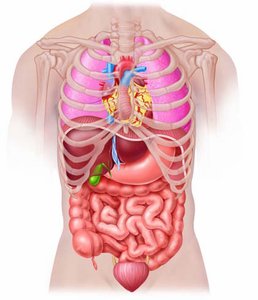
Ventral body cavity: Subdivided into the thoracic cavity (heart and lungs) and the abdominopelvic cavity (digestive, urinary, and reproductive organs).
Serous Membranes
The ventral body cavity and its organs are lined with serous membranes (serosae):
Parietal serosa: Lines the cavity walls.
Visceral serosa: Covers the organs.
Serous fluid between layers reduces friction and compartmentalizes organs to prevent infection spread.
Specific names: Peritoneum (abdominal), Pleura (lungs), Pericardium (heart).
Abdominopelvic Quadrants and Regions
For clinical and anatomical purposes, the abdominopelvic cavity is divided into:
Four quadrants: Right upper (RUQ), right lower (RLQ), left upper (LUQ), left lower (LLQ).
Nine regions: Umbilical, epigastric, hypogastric (pubic), right/left iliac (inguinal), right/left lumbar, right/left hypochondriac.

Other Body Cavities
Smaller Body Cavities
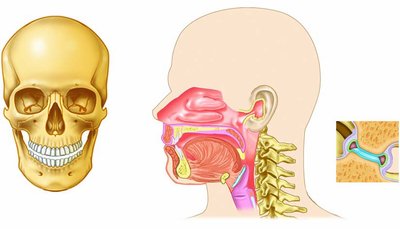
In addition to the major cavities, the body contains several smaller cavities, mainly in the head:
Oral cavity: Mouth, contains teeth and tongue, continuous with the digestive tract.
Nasal cavity: Part of the respiratory system, located within and posterior to the nose.
Orbital cavities: House the eyes.
Middle ear cavities: Contain bones that transmit sound vibrations.
Synovial cavities: Found in freely movable joints, lined with lubricating fluid-secreting membranes.
Summary Table: Major Body Cavities and Contents
Body Cavity | Location | Main Organs | Protection |
|---|---|---|---|
Cranial (Dorsal) | Within skull | Brain | High (bone) |
Vertebral (Dorsal) | Within vertebral column | Spinal cord | High (bone) |
Thoracic (Ventral) | Chest, above diaphragm | Heart, lungs | Moderate (rib cage) |
Abdominal (Ventral) | Below diaphragm, above pelvis | Stomach, intestines, liver, etc. | Low (muscle, fat) |
Pelvic (Ventral) | Within pelvis | Bladder, reproductive organs, rectum | Moderate (pelvic bones) |
Key Terms and Definitions
Anatomical position: Standard body orientation for reference.
Directional terms: Words describing location of structures (e.g., superior, inferior, anterior, posterior, medial, lateral, proximal, distal, superficial, deep).
Body planes: Imaginary lines dividing the body (sagittal, frontal, transverse).
Body cavities: Spaces within the body housing organs (dorsal, ventral, cranial, vertebral, thoracic, abdominal, pelvic).
Serous membranes: Double-layered membranes lining ventral body cavities and organs.
Practice and Application
Practice identifying body regions, planes, and cavities on anatomical models and diagrams.
Use correct terminology to describe the location of organs and structures relative to each other.
Apply knowledge of planes and sections to interpret medical imaging (e.g., MRI, CT scans).
