 뒤로
뒤로The Neuron: Structure, Function, and Membrane Properties
스터디 가이드 - 스마트 노트
 자료에 맞춘 맞춤형 노트, 핵심 정의, 예시, 맥락을 확장해 제공합니다.
The Neuron: Structure and Function
Overview of Neurons
Neurons are the fundamental excitable cells of the nervous system, specialized for the rapid transmission of electrical and chemical signals. They communicate with each other and with other cell types through synaptic transmission, enabling complex processes such as sensation, movement, and cognition.
Excitability: Neurons can generate and propagate action potentials in response to stimuli.
Synaptic Transmission: Communication between neurons occurs at specialized junctions called synapses.
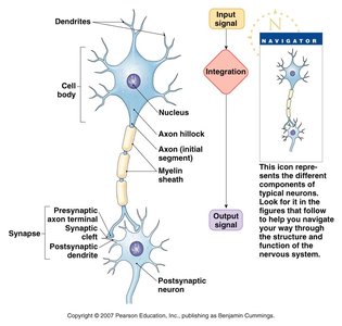
Cell Body (Soma): Contains the nucleus and metabolic machinery of the cell.
Dendrites: Receive input signals from other neurons.
Axon: Conducts action potentials away from the cell body toward synaptic terminals.
Neuron Morphologies
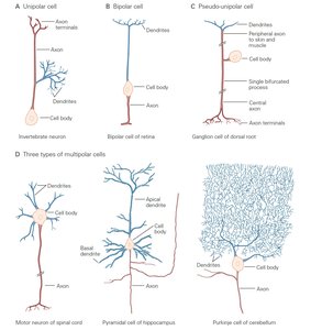
Neurons exhibit a variety of morphologies, each adapted to specific functional roles within the nervous system. The main types include unipolar, bipolar, pseudo-unipolar, and multipolar neurons.
Unipolar: Single process extends from the cell body (common in invertebrates).
Bipolar: Two processes (one axon, one dendrite); found in sensory organs like the retina.
Pseudo-unipolar: Single process that splits into two branches; typical of sensory neurons in dorsal root ganglia.
Multipolar: Multiple dendrites and one axon; most common type in the CNS (e.g., motor neurons, Purkinje cells).
Glial Cells
Types and Functions of Glial Cells
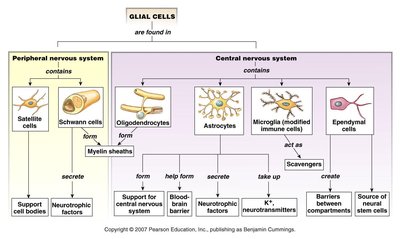
Glial cells are non-neuronal cells that provide structural and functional support to neurons. They are essential for maintaining homeostasis, forming myelin, and participating in signal transmission in the nervous system.
Peripheral Nervous System (PNS):
Satellite cells: Support cell bodies.
Schwann cells: Form myelin sheaths around axons.
Central Nervous System (CNS):
Oligodendrocytes: Form myelin sheaths.
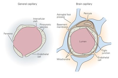
Astrocytes: Support, form blood-brain barrier, secrete neurotrophic factors, regulate neurotransmitters.
Microglia: Immune cells, act as scavengers.
Ependymal cells: Create barriers between compartments, source of neural stem cells.
Electrical Properties of Neurons
Basic Electrical Concepts
Neurons are electrically excitable due to the presence of ion channels in their membranes. The key electrical properties include voltage (V), current (I), resistance (R), and conductance (G).
Voltage (V): Difference in electrical potential across the membrane (measured in mV).
Current (I): Flow of electrical charge (measured in pA or nA).
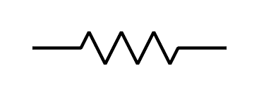
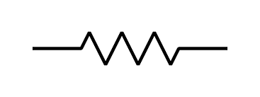
Resistance (R): Opposition to current flow (measured in MΩ); inverse is conductance (G, in nS).
Ohm’s Law:
or
Membrane Capacitance and Time Constant
The lipid bilayer of the neuron acts as a capacitor, storing and separating charge. Capacitance (C) determines how quickly the membrane potential (Vm) can change in response to current.
Capacitance (C): Amount of charge needed to change the membrane potential by a certain amount (measured in pF).
Membrane Time Constant (τm): Time required for Vm to reach 63% of its final value after a current step.
To speed up changes in Vm, decrease membrane capacitance (Cm) or resistance (Rm).
Membrane Potential and Ion Channels
Neurons maintain a resting membrane potential (RMP) of approximately -70 mV, primarily due to the selective permeability of the membrane to different ions and the activity of ion pumps.
Depolarization: Vm becomes more positive (e.g., influx of Na+).
Hyperpolarization: Vm becomes more negative (e.g., efflux of K+ or influx of Cl-).
Equilibrium Potentials and the Nernst Equation
Equilibrium Potential for a Single Ion
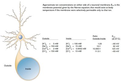
The equilibrium potential (Eion) is the membrane voltage at which the net flow of a particular ion across the membrane is zero. It is determined by the Nernst equation:
R = universal gas constant
T = temperature (Kelvin)
n = ion valence
F = Faraday’s constant
[i] = ion concentration
Resting Membrane Potential with Multiple Ions
When the membrane is permeable to more than one ion, the resting membrane potential (Vm) is a weighted average of the equilibrium potentials for all permeant ions, weighted by their conductances (g):
Maintenance of Ionic Gradients
The Na+-K+ Pump
The Na+-K+ ATPase actively transports 3 Na+ ions out of the cell and 2 K+ ions into the cell, maintaining the concentration gradients necessary for the resting membrane potential. This pump consumes a significant portion of the brain’s metabolic energy.
Regulation of the Ionic Environment
Role of the Blood-Brain Barrier, Renal System, and Glia
The ionic environment of neurons is tightly regulated by several mechanisms:
Blood-Brain Barrier: Restricts movement of ions and molecules between blood and brain extracellular fluid.
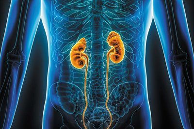
Renal System: Maintains systemic ionic balance.
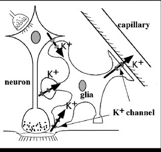
Glial Cells: Regulate extracellular ion concentrations, especially K+.
Consequences of Ionic Dysregulation
Abnormal extracellular ion concentrations can lead to changes in neuronal excitability:
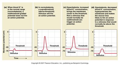
Hyperkalemia: Elevated extracellular K+ depolarizes neurons, increasing excitability and risk of spontaneous action potentials.
Hypokalemia: Reduced extracellular K+ hyperpolarizes neurons, decreasing excitability.
Summary Table: Key Ion Concentrations and Equilibrium Potentials
Ion | Outside (mM) | Inside (mM) | Ratio (Out/In) | Eion (mV, 37°C) |
|---|---|---|---|---|
K+ | 5 | 150 | 1:30 | -90 |
Na+ | 150 | 15 | 10:1 | +60 |
Cl- | 120 | 10 | 12:1 | -65 |
Ca2+ | 2 | 0.0002 | 10,000:1 | +122 |
Key Equations
Ohm’s Law: or
Membrane Time Constant:
Nernst Equation:
Resting Membrane Potential (multiple ions):
Additional info: This guide covers the structure and function of neurons, the role of glial cells, the electrical properties of neuronal membranes, and the physiological basis of the resting membrane potential. It also addresses the importance of ionic regulation for neuronal excitability and the consequences of dysregulation.
