 뒤로
뒤로The Organization of the Human Body: Body Cavities, Abdominopelvic Regions, and Serous Membranes
스터디 가이드 - 스마트 노트
 자료에 맞춘 맞춤형 노트, 핵심 정의, 예시, 맥락을 확장해 제공합니다.
Module 1.4: The Organization of the Human Body
Body Cavities of the Human Body
The human body contains several major cavities that protect internal organs and allow them to move and expand. These cavities are divided into two main groups: the posterior (dorsal) body cavity and the anterior (ventral) body cavity.
Posterior Body Cavity: Located on the back (posterior) side of the body, it includes:
Cranial cavity: Found within the skull, it protects the brain.
Vertebral (spinal) cavity: Located within the vertebral column, it protects the spinal cord. Both cavities are continuous and filled with cerebrospinal fluid (CSF), which bathes and supports the brain and spinal cord.
Anterior Body Cavity: Located on the front (anterior) side of the body, it is divided by the diaphragm muscle into:
Thoracic cavity: Superior to the diaphragm, contains the lungs and heart.
Abdominopelvic cavity: Inferior to the diaphragm, contains digestive, urinary, and reproductive organs.
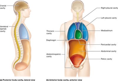
Subdivisions of the Anterior Body Cavity
Thoracic Cavity:
Pleural cavities: Right and left, each surrounds a lung and is lined by serous membranes.
Mediastinum: Central compartment between the pleural cavities; contains the heart, great blood vessels, trachea, and esophagus. The mediastinum itself is not within a serous membrane.
Pericardial cavity: Located within the mediastinum, surrounds the heart and is lined by serous membranes.
Abdominopelvic Cavity:
Abdominal cavity: Extends from the diaphragm to the bony pelvis; contains digestive, lymphatic, and urinary organs. Includes the peritoneal cavity (a serous membrane-lined subcavity).
Pelvic cavity: Enclosed by the bony pelvis; contains reproductive, digestive, and urinary organs.
Abdominopelvic Quadrants and Regions
The abdominopelvic cavity can be divided for clinical and anatomical purposes:
Four Quadrants: Created by a transverse and a midsagittal line through the umbilicus:
Right Upper Quadrant (RUQ)
Left Upper Quadrant (LUQ)
Right Lower Quadrant (RLQ)
Left Lower Quadrant (LLQ)
Nine Regions: Created by two parasagittal and two transverse lines:
Right/Left Hypochondriac Regions (superior, under ribs)
Epigastric Region (superior, above stomach)
Right/Left Lumbar Regions (middle, lateral to umbilicus)
Umbilical Region (center, contains umbilicus)
Right/Left Iliac (Inguinal) Regions (inferior, over iliac crests)
Hypogastric Region (inferior, below stomach)
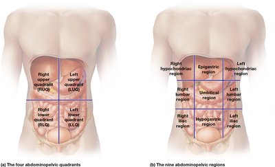
Clinical Application: The quadrant system is commonly used in medicine to localize abdominal pain and narrow down possible causes based on the organs present in each quadrant (e.g., RLQ pain may indicate appendicitis).
Serous Membranes and Serous Cavities
Serous membranes are thin sheets of tissue that line certain body cavities and cover many organs. They consist of a single, continuous layer that folds over itself, creating two layers:
Visceral layer: Contacts the organ directly.
Parietal layer: Contacts surrounding structures.
Serous fluid: A thin, watery fluid between the layers that lubricates and reduces friction as organs move.
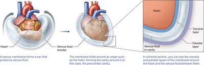
Major serous membranes and their associated cavities include:
Pleural membranes: Surround the lungs (pleural cavities).
Pericardial membranes: Surround the heart (pericardial cavity).
Peritoneal membranes: Surround many abdominal organs (peritoneal cavity). Organs within the peritoneal cavity are called intraperitoneal; those behind it are retroperitoneal (e.g., kidneys).
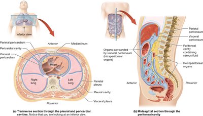
Clinical Note: Inadequate serous fluid (e.g., in pleural cavities) increases friction and can impair organ function.
Medical Imaging of Body Cavities
Medical imaging techniques allow visualization of internal body structures and cavities without surgery. Common modalities include:
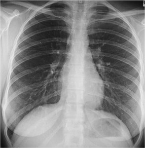
X-ray: Uses ionizing radiation to produce images of dense structures (e.g., bones, thoracic cavity).
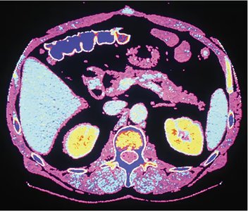
Computed Tomography (CT) Scan: Uses ionizing radiation and computer processing to create detailed cross-sectional images, often in the transverse plane.
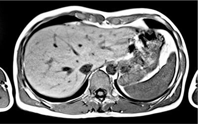
Magnetic Resonance Imaging (MRI): Uses magnetic fields and computer processing to produce high-resolution images, especially useful for soft tissues.
These imaging techniques are essential for diagnosing diseases, planning surgeries, and monitoring treatment outcomes.
