 뒤로
뒤로The Plasma Membrane and Membrane Potential: Structure, Function, and Transport
스터디 가이드 - 스마트 노트
 자료에 맞춘 맞춤형 노트, 핵심 정의, 예시, 맥락을 확장해 제공합니다.
The Plasma Membrane and Membrane Potential
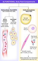
The Three Fluid Compartments
The human body contains three main fluid compartments that are essential for physiological function. These compartments are separated by biological membranes, which regulate the movement of substances between them.
Extracellular Fluid (ECF): Fluid outside the cells, further divided into plasma (the fluid portion of blood) and interstitial fluid (fluid that surrounds most cells).
Intracellular Fluid (ICF): Fluid within the cells, making up the majority of total body water.
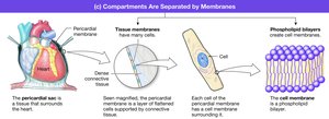
Compartments Are Separated by Membranes
Biological membranes serve as barriers that separate the body's fluid compartments. These membranes are composed of phospholipid bilayers and associated proteins, providing both structural integrity and selective permeability.
Tissue membranes: Consist of layers of cells and connective tissue, such as the pericardial sac around the heart.
Cell membranes: Phospholipid bilayers that enclose individual cells, maintaining the internal environment.
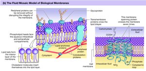
Biological Membranes: Structure and Composition
The Fluid Mosaic Model
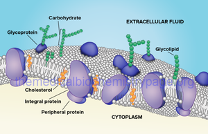
The plasma membrane is described by the fluid mosaic model, which depicts the membrane as a dynamic structure with proteins floating in or on a fluid lipid bilayer. This model explains the flexibility and varied functions of the membrane.
Lipids: Primarily phospholipids, which are amphipathic molecules with hydrophilic heads and hydrophobic tails, forming a bilayer that acts as a barrier to water-soluble substances.
Cholesterol: Interspersed within the bilayer, cholesterol maintains membrane fluidity and stability by preventing fatty acid tails from packing too closely.
Proteins: Integral and peripheral proteins serve as channels, carriers, receptors, enzymes, and cell adhesion molecules (CAMs).
Carbohydrates: Attached to proteins (glycoproteins) or lipids (glycolipids) on the extracellular surface, these molecules are involved in cell recognition and signaling.
Function of the Lipid Bilayer
The lipid bilayer forms the fundamental structure of the plasma membrane, providing a hydrophobic barrier that restricts the passage of water-soluble substances and contributes to membrane fluidity.
Barrier function: Prevents free movement of ions and polar molecules between the ICF and ECF.
Fluidity: Allows for membrane flexibility and the movement of embedded proteins.
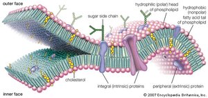
Carbohydrates in the Plasma Membrane
Carbohydrates are found on the outer surface of the plasma membrane, attached to proteins and lipids. They play a crucial role in cell recognition, immune response, and tissue organization.
Glycoproteins and glycolipids: Serve as markers for cellular identification and communication.
Self-recognition: Helps the immune system distinguish self from non-self cells.
Cell-to-Cell Adhesion and Specialized Junctions
Cell-to-Cell Adhesion
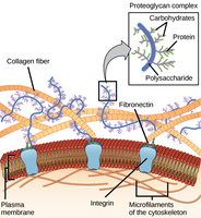
Cells are bound together to form tissues and organs through various adhesion mechanisms, including the extracellular matrix and cell adhesion molecules (CAMs).
Extracellular matrix: A network of fibrous proteins and polysaccharides that provides structural support and mediates cell signaling.
Cell adhesion molecules (CAMs): Membrane proteins that connect cells via hooks and loops, facilitating tissue integrity.
Specialized Cell Junctions
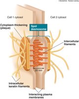
Specialized junctions provide mechanical strength and regulate communication between adjacent cells.
Desmosomes: Adhering junctions that provide strong attachments between cells, especially in tissues subject to stretching (e.g., skin, heart).
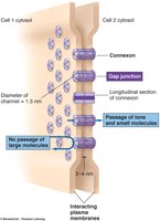
Gap junctions: Channels that connect the cytoplasm of adjacent cells, allowing the passage of ions and small molecules for intercellular communication.
Membrane Transport Mechanisms
Selective Permeability of the Plasma Membrane
The plasma membrane is selectively permeable, allowing certain substances to cross while restricting others. Permeability depends on solubility, size, and the presence of specific transport mechanisms.
Nonpolar and small molecules: Can diffuse freely across the membrane.
Polar, charged, or large molecules: Require assistance from membrane proteins.
Unassisted Membrane Transport
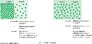
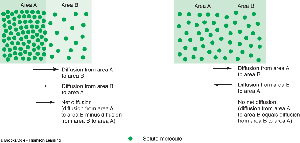
Some molecules can cross the membrane without assistance, primarily through diffusion down their concentration gradients.
Simple diffusion: Movement of molecules from an area of higher concentration to lower concentration due to random thermal motion.
Equilibrium: Achieved when the concentration of the substance is equal on both sides of the membrane.
Fick’s Law of Diffusion
The rate of diffusion across a membrane is described by Fick’s Law, which considers several factors:
Increases rate: Greater concentration gradient (ΔC), higher membrane permeability (P), larger surface area (A).
Decreases rate: Higher molecular weight (MW), greater distance (ΔX).
Fick's Law equation:
Assisted Membrane Transport
Large, polar, or charged molecules require assistance to cross the plasma membrane. This is achieved through protein-mediated mechanisms:
Facilitated diffusion: Movement down a concentration gradient via channel or carrier proteins, without energy input.
Osmosis: Diffusion of water through aquaporins.
Active transport: Movement against a concentration gradient, requiring energy (usually ATP) and carrier proteins.
Vesicular transport: Bulk movement of materials via endocytosis or exocytosis.
Channel Proteins
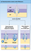
Channel proteins form water-filled passageways that allow specific ions or water molecules to cross the membrane. They can be classified as open (leaky) or gated channels, which open or close in response to signals.
Types of gated channels: Chemically gated, voltage-gated, and mechanically gated.
Carrier Proteins
Carrier proteins bind to specific molecules and undergo conformational changes to transport them across the membrane. They can function as uniporters (one substance), symporters (two substances in the same direction), or antiporters (two substances in opposite directions).
Facilitated diffusion: Down the concentration gradient, no energy required.
Active transport: Against the concentration gradient, requires energy.
Osmosis and Osmolarity
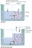
Osmosis is the movement of water across a selectively permeable membrane from an area of lower solute concentration to higher solute concentration. Osmolarity refers to the total concentration of solute particles in a solution, and osmotic pressure is the force generated by water movement.
Facilitated Diffusion vs. Active Transport
Facilitated diffusion: Down concentration or electrical gradient, uses channels or carriers, no energy required.
Active transport: Against concentration gradient, uses carrier proteins, requires energy (e.g., Na+/K+ pump).
Membrane Potential
The Resting Membrane Potential
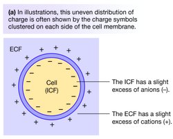
Cells maintain a resting membrane potential due to the unequal distribution of ions across the plasma membrane and the selective permeability of the membrane. The inside of the cell (ICF) is typically more negative than the outside (ECF).
ICF: Net negative charge (excess anions).
ECF: Net positive charge (excess cations).
Ion Concentrations and Permeability
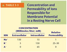
The resting membrane potential is primarily determined by the concentrations and relative permeabilities of sodium (Na+), potassium (K+), and anions (A-).
Ion | Extracellular (mM) | Intracellular (mM) | Relative Permeability |
|---|---|---|---|
Na+ | 150 | 15 | 1 |
K+ | 5 | 150 | 25–30 |
A- | 0 | 65 | 0 |
Equilibrium Potential and the Nernst Equation
The equilibrium potential for an ion is the membrane potential at which there is no net movement of that ion across the membrane. It can be calculated using the Nernst equation:
Example for K+:
Example for Na+:
Goldman Equation
The Goldman equation calculates the resting membrane potential (Vm) by considering the relative permeabilities and concentrations of multiple ions (mainly Na+ and K+):
PK: Permeability of K+ (set as 1)
PNa: Permeability of Na+ (typically 0.04)
Net Force on Ions
The net force acting on an ion is the difference between the membrane potential (Vm) and the equilibrium potential (Ex) for that ion:
If positive, the force drives a positive ion out of the cell.
If negative, the force drives a positive ion into the cell.
If zero, there is no net force.
Physiological Importance of Membrane Potential
Resting membrane potential is essential for the function of nerve and muscle cells, enabling rapid changes in membrane potential that underlie nerve impulses and muscle contraction.
Additional info: The Na+/K+ pump actively maintains the concentration gradients of Na+ and K+, which are critical for the generation and maintenance of the resting membrane potential.
