 뒤로
뒤로The Special Sense of Vision: Anatomy and Physiology of the Eye
스터디 가이드 - 스마트 노트
 자료에 맞춘 맞춤형 노트, 핵심 정의, 예시, 맥락을 확장해 제공합니다.
The Special Sense of Vision
Introduction
The human visual system is a complex network that allows us to perceive and interpret our environment through the detection of light. This section explores the anatomy of the eye, the physiology of vision, and the neural pathways involved in processing visual information.
Anatomy of the Eye
Internal Structure of the Eye
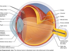
The eye is a nearly spherical organ composed of three layers and several internal structures that contribute to its function as a sensory receptor for light.
Sclera: The tough, white outer layer that maintains the shape of the eye.
Choroid: Vascular layer providing oxygen and nutrients to the eye.
Retina: The innermost layer containing photoreceptors (rods and cones).
Cornea: Transparent anterior portion that refracts light.
Lens: Biconvex structure that focuses light onto the retina.
Aqueous humor: Fluid in the anterior segment, maintaining intraocular pressure.
Vitreous humor: Gel-like substance in the posterior segment, supporting the retina.
Optic nerve: Transmits visual information to the brain.
Light and Optics
Wavelength and Color
Visible light is a small portion of the electromagnetic spectrum, with wavelengths between 400-700 nm. The color perceived by the eye depends on the wavelength of light reflected by objects. For example, grass appears green because it reflects green wavelengths and absorbs others.
Red: Longest wavelength, lowest energy.
Violet: Shortest wavelength, highest energy.
White: Reflects all colors.
Black: Absorbs all colors.
Refraction and Lenses
Refraction is the bending of light rays as they pass from one medium to another at an oblique angle. The eye uses convex lenses to converge light rays at a focal point on the retina, forming an inverted and reversed image. Concave lenses disperse light and are used to correct certain vision problems.
Focusing Light on the Retina
Pathway of Light
Light passes through the following structures before reaching the photoreceptors:
Cornea
Aqueous humor
Lens
Vitreous humor
Neural layer of retina
Photoreceptors
Light is refracted three times: entering the cornea, entering the lens, and leaving the lens. The lens changes curvature for fine focusing.
Focusing for Distant and Close Vision
The eye adjusts its focusing power depending on the distance of the object being viewed.
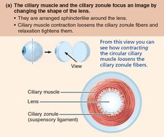
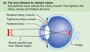
Distant Vision: The ciliary muscles relax, the ciliary zonule tightens, and the lens flattens to focus parallel rays from distant objects.
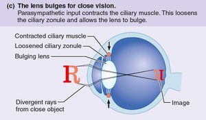
Close Vision: The ciliary muscles contract, the ciliary zonule loosens, and the lens bulges to increase refraction for divergent rays from close objects.
Accommodation, Pupil Constriction, and Convergence
Accommodation: Changing lens shape to focus on near objects. The closest point the eye can focus is the near point of vision.
Pupil Constriction: Reduces divergent light rays entering the eye, mediated by the parasympathetic nervous system.
Convergence: Medial rotation of the eyeballs to maintain focus on a close object.
Clinical Correlates: Problems of Refraction
Errors in refraction are often due to the shape of the eyeball or lens.
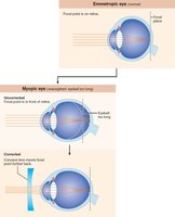
Myopia (Nearsightedness): Eyeball too long; focal point in front of retina. Corrected with concave lenses.
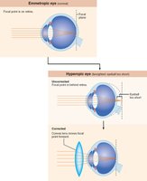
Hyperopia (Farsightedness): Eyeball too short; focal point behind retina. Corrected with convex lenses.
Astigmatism: Unequal curvature of cornea or lens; corrected with cylindrical lenses or laser procedures.
Retina and Photoreceptors
Structure of the Retina
The retina consists of two layers:
Pigmented Layer: Absorbs light, prevents scattering, stores vitamin A, and phagocytizes cell fragments.
Neural Layer: Contains photoreceptors (rods and cones), bipolar cells, and ganglion cells. Signals pass from photoreceptors to bipolar cells to ganglion cells, whose axons form the optic nerve.
Photoreceptors: Rods and Cones
Photoreceptors are specialized neurons that detect light.
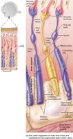
Rods: More numerous, highly sensitive to dim light, responsible for night and peripheral vision, but do not detect color or sharp images.
Cones: Less sensitive, require bright light, responsible for high-resolution color vision. Concentrated in the macula lutea and fovea centralis.
Functional Anatomy of Photoreceptors
Outer segments contain visual pigments that change shape when absorbing light.
Inner segments connect to the cell body.
Photoreceptors are vulnerable to damage and are renewed every 24 hours.
Visual Pigments and Phototransduction
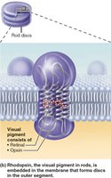
Visual pigments consist of retinal (derived from vitamin A) and opsins. Rods contain rhodopsin; cones contain one of three opsins (red, green, blue).
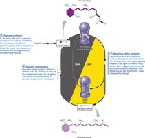
Retinal exists in two isomers: 11-cis (bent, in the dark) and all-trans (straight, after absorbing light).
Conversion from 11-cis to all-trans triggers a cascade leading to electrical impulses.
Phototransduction: Capturing Light
Phototransduction is the process by which light energy is converted into a graded receptor potential.
Pigment Synthesis: Opsin and 11-cis retinal combine to form rhodopsin in the dark.
Pigment Bleaching: Light converts 11-cis retinal to all-trans, causing rhodopsin to break down.
Pigment Regeneration: All-trans retinal is converted back to 11-cis, regenerating rhodopsin.
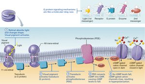
Phototransduction Mechanism
Light-activated rhodopsin triggers a G protein (transducin), which activates phosphodiesterase (PDE) to break down cyclic GMP (cGMP). In darkness, cGMP keeps cation channels open, depolarizing the cell. In light, cGMP decreases, channels close, and the cell hyperpolarizes, signaling vision.
Signal Transmission in the Retina
Information Processing
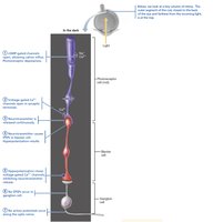
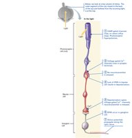
Photoreceptors and bipolar cells generate graded potentials. In darkness, photoreceptors release glutamate, inhibiting bipolar cells. In light, hyperpolarization stops glutamate release, allowing bipolar cells to depolarize and stimulate ganglion cells, which generate action potentials sent to the brain.
Light and Dark Adaptation
Dark Adaptation: Moving from bright to dark, rods regenerate rhodopsin, sensitivity increases over 20–30 minutes, and pupils dilate.
Light Adaptation: Moving from dark to bright, rods and cones are strongly stimulated, pigments break down, pupils constrict, and visual acuity improves over 5–10 minutes as cones adapt.
Clinical Correlates of Phototransduction
Nyctalopia (Night Blindness): Impaired rod function, often due to vitamin A deficiency or degenerative diseases like retinitis pigmentosa.
Neural Pathways and Visual Processing
Visual Pathway to the Brain
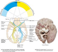
Axons of retinal ganglion cells form the optic nerve. At the optic chiasma, medial fibers decussate. Most fibers synapse in the lateral geniculate nucleus of the thalamus, then project via optic radiations to the primary visual cortex in the occipital lobe. Some fibers project to the midbrain for visual reflexes and to the hypothalamus for circadian rhythms.
Cortical Processing
Occipital lobe: Processes form, color, and movement.
Ventral stream ("What"): Identifies objects.
Dorsal stream ("Where"): Assesses spatial location.
Both streams contribute to motor responses via the frontal cortex.
Depth Perception
Depth perception arises from the fusion of slightly different images from each eye in the visual cortex, resulting in three-dimensional vision. This process requires input from both eyes.
