 뒤로
뒤로The Special Senses: Olfaction, Gustation, Vision, and Hearing
스터디 가이드 - 스마트 노트
 자료에 맞춘 맞춤형 노트, 핵심 정의, 예시, 맥락을 확장해 제공합니다.
The Special Senses
Introduction to the Special Senses
The special senses include olfaction (smell), gustation (taste), vision, equilibrium (balance), and hearing. These senses are mediated by specialized organs and pathways that allow the body to detect and interpret environmental stimuli.
Olfaction (Smell)
Olfactory Organs and Structure
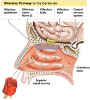
Olfaction is the sense of smell, mediated by olfactory organs located in the nasal cavity. These organs consist of two main layers:
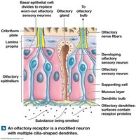
Olfactory epithelium: Contains olfactory sensory neurons, supporting cells, and basal epithelial cells (stem cells).
Lamina propria: Underlies the epithelium and contains areolar tissue, blood vessels, nerves, and olfactory glands that secrete mucus.
Olfactory Receptors and Pathways
Odorants are small, airborne, organic molecules that stimulate olfactory receptors.
Olfactory sensory neurons have dendrites that extend into the mucus, where odorant-binding proteins are located.
The axons of these neurons form the olfactory nerves, which pass through the cribriform plate to synapse in the olfactory bulbs.
From the olfactory bulbs, information is relayed via the olfactory tracts to the olfactory cortex, hypothalamus, and limbic system.
Olfactory information is unique in that it reaches the cerebral cortex without first passing through the thalamus.
Physiology of Olfaction
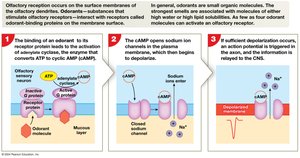
Odorant molecules bind to G protein–coupled receptors on olfactory dendrites.
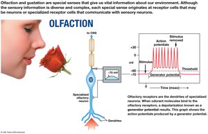
This activates a second messenger (cAMP), which opens sodium ion channels, depolarizing the neuron and generating a potential.
If the depolarization is sufficient, an action potential is triggered and transmitted to the CNS.
Olfactory Discrimination and Adaptation
Humans can distinguish between 2,000–4,000 odorants, with more than 50 primary smells interpreted by patterns of receptor activity.
Olfactory neurons are replaced frequently, but their number declines with age.
Olfactory adaptation occurs centrally, with other brain nuclei modulating the olfactory bulbs.
Gustation (Taste)
Gustatory Organs and Papillae
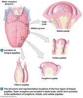
Gustation is the sense of taste, providing information about substances consumed. Taste receptor cells are found in taste buds, which are distributed on the tongue and parts of the pharynx and larynx. The tongue contains several types of papillae:
Filiform papillae: Provide friction, do not contain taste buds.
Fungiform papillae: Contain about five taste buds each, located mainly at the tip and sides of the tongue.
Vallate papillae: Large, arranged in a V-shape at the posterior tongue, each with about 100 taste buds.
Foliate papillae: Found on the lateral margins, contain taste buds.
Taste Bud Structure
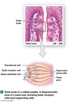
Taste buds contain basal epithelial cells (stem cells), transitional cells (supporting cells), and taste receptor cells (gustatory epithelial cells).
Taste receptor cells extend microvilli (taste hairs) into the taste pore, where they interact with dissolved substances.
These cells synapse with sensory neurons.
Gustatory Pathways
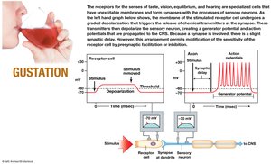
Activation of taste receptor cells leads to neurotransmitter release, stimulating sensory neurons.
The facial nerve (VII) innervates the anterior two-thirds of the tongue, the glossopharyngeal nerve (IX) innervates the vallate papillae, and the vagus nerve (X) innervates extralingual taste buds.
Sensory fibers synapse in the solitary nucleus of the medulla, then ascend to the thalamus and finally to the gustatory cortex in the insula.
Taste perception is influenced by texture (somatosensory input) and olfactory information.
Physiology of Taste
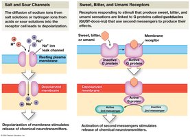
Salty and sour tastes are detected by the diffusion of ions (Na+ for salty, H+ for sour) through leak channels, leading to depolarization.
Sweet, bitter, and umami tastes are detected by G protein–coupled receptors, which activate second messenger pathways.
Gustatory Discrimination
Primary taste sensations: sweet, salty, sour, bitter, umami, and water.
Umami is a savory taste detected by glutamate receptors; water is detected by receptors in the pharynx.
Humans are more sensitive to bitter and sour tastes, which may serve as a protective mechanism against toxins.
Taste sensitivity varies among individuals and decreases with age.
Vision
Accessory Structures of the Eye
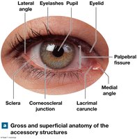
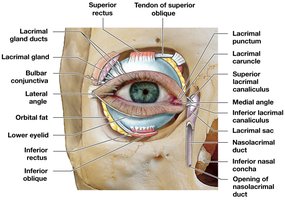
The eye is protected and supported by accessory structures, including the eyelids, eyelashes, conjunctiva, and the lacrimal apparatus.
Eyelids (palpebrae): Protect and lubricate the eye; contain tarsal glands that secrete oils to prevent sticking.
Eyelashes: Prevent debris from entering the eye.
Conjunctiva: Epithelial membrane covering the inner eyelids and anterior eye surface.
Lacrimal apparatus: Produces and drains tears, which clean, moisten, and protect the eye.
Anatomy of the Eyeball
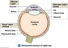
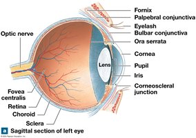
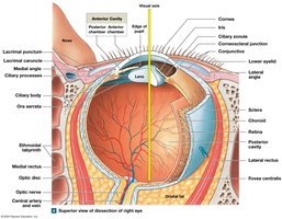
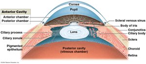
The eyeball is a hollow sphere with three layers: fibrous (sclera and cornea), vascular (uvea: iris, ciliary body, choroid), and inner (retina).
It contains two cavities: anterior (aqueous humor) and posterior (vitreous body).
Fibrous Layer
Sclera: The white, tough outer layer providing protection and shape.
Cornea: Transparent anterior part allowing light entry; avascular and easily damaged.
Corneoscleral junction (limbus): Border between cornea and sclera.
Vascular Layer (Uvea)
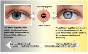
Iris: Colored part, controls pupil size via sphincter and dilator muscles.
Ciliary body: Contains ciliary muscle and processes; controls lens shape via the ciliary zonule (suspensory ligament).
Choroid: Vascular, pigmented layer supplying nutrients to the retina.
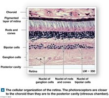
Inner Layer (Retina)
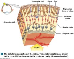
Pigmented layer: Absorbs stray light, supports photoreceptors.
Neural layer: Contains photoreceptors (rods and cones), bipolar cells, ganglion cells, horizontal and amacrine cells.
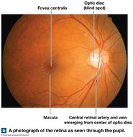
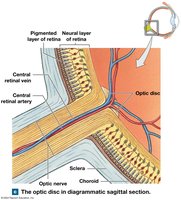
Macula: Area of sharpest vision, with the fovea centralis at its center.
Optic disc: Blind spot where the optic nerve exits; no photoreceptors present.
Fluids of the Eye
Aqueous humor: Circulates in the anterior cavity, provides nutrients, maintains intraocular pressure, drains via the scleral venous sinus.
Vitreous body: Gelatinous mass in the posterior cavity, stabilizes eye shape.
Lens and Accommodation
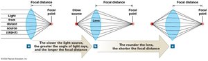
Lens: Transparent, biconvex, focuses light on the retina by changing shape (accommodation).
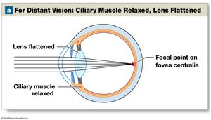
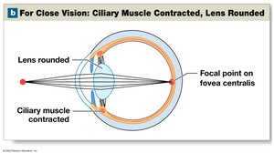
Accommodation: Lens rounds for near vision (ciliary muscle contracts), flattens for distant vision (ciliary muscle relaxes).
Cataracts: Loss of lens transparency, often age-related.
Formation of a Visual Image
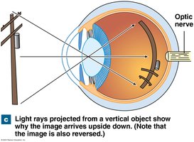
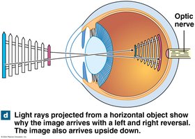
Light is refracted by the cornea and lens to focus on the retina.
The image is inverted and reversed; the brain corrects this orientation.
Visual acuity is the clarity of vision, with 20/20 as the standard.
Common refractive errors: myopia (nearsightedness), hyperopia (farsightedness), astigmatism (irregular curvature).
Physiology of Vision
Photoreceptors
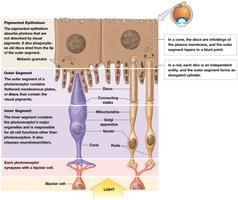
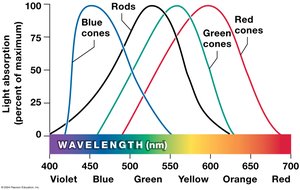
Rods: Sensitive to low light, do not detect color.
Cones: Detect color (blue, green, red cones), require more light, concentrated in the fovea.
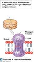
Both have inner and outer segments; outer segments contain visual pigments.
Phototransduction
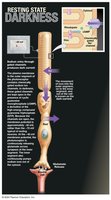
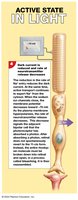
In darkness, rods are depolarized due to open sodium channels (maintained by cGMP), releasing glutamate.
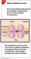
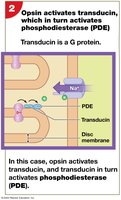
Light absorption changes retinal from 11-cis to 11-trans form, activating opsin, which activates transducin and phosphodiesterase (PDE).
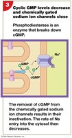
PDE breaks down cGMP, closing sodium channels, hyperpolarizing the cell, and reducing neurotransmitter release.
The Ear: Hearing and Equilibrium
Anatomy of the Ear
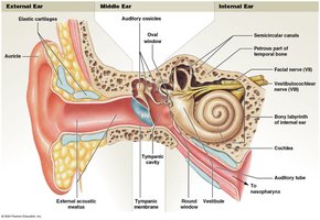
The ear is divided into three regions: external, middle, and internal ear. Each region has specialized structures for hearing and equilibrium.
External ear: Auricle (pinna), external acoustic meatus, tympanic membrane.
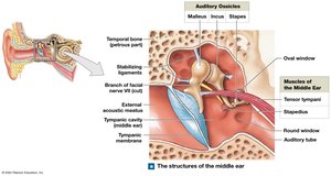
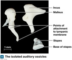
Middle ear: Tympanic cavity, auditory ossicles (malleus, incus, stapes), auditory tube.
Internal ear: Bony and membranous labyrinths, vestibule, semicircular canals, cochlea.
Middle Ear and Auditory Ossicles
Ossicles transmit and amplify vibrations from the tympanic membrane to the oval window of the internal ear.
Muscles (tensor tympani and stapedius) protect the ear from loud sounds by dampening ossicle movement.
Internal Ear: Labyrinths and Sensory Regions
Bony labyrinth: Contains perilymph, surrounds the membranous labyrinth.
Membranous labyrinth: Contains endolymph, houses sensory receptors.
Divided into vestibule (saccule and utricle for equilibrium), semicircular canals (rotational movement), and cochlea (hearing).
Equilibrium
Hair cells in the vestibular complex detect head position and movement.
Semicircular ducts detect rotational movement; utricle and saccule detect linear acceleration and gravity.
Hair cells have stereocilia and a kinocilium; bending toward the kinocilium depolarizes the cell, away hyperpolarizes it.
Hearing
Sound waves vibrate the tympanic membrane, transmitted via ossicles to the oval window, creating pressure waves in the cochlear perilymph.
Pressure waves move the basilar membrane, stimulating hair cells in the spiral organ (organ of Corti).
Hair cell depolarization leads to neurotransmitter release and stimulation of sensory neurons in the cochlear nerve.
Sound Properties
Frequency (Hz): Determines pitch; high frequency = high pitch.
Amplitude: Determines loudness (measured in decibels, dB).
Cochlear Structure and Function
Cochlear duct (scala media) is filled with endolymph, lies between scala vestibuli and scala tympani (perilymph).
Spiral organ rests on the basilar membrane; hair cells contact the tectorial membrane.
High-frequency sounds vibrate the basilar membrane near the oval window; low-frequency sounds travel further along the cochlea.
Summary Table: Comparison of Special Senses
Sense | Receptor Type | Location | Main Pathway |
|---|---|---|---|
Olfaction | Olfactory neurons | Nasal cavity | Olfactory bulb → tract → cortex |
Gustation | Taste receptor cells | Taste buds (tongue, pharynx, larynx) | Cranial nerves VII, IX, X → medulla → thalamus → cortex |
Vision | Rods and cones | Retina | Optic nerve → thalamus → visual cortex |
Hearing | Hair cells | Cochlea | Cochlear nerve → brainstem → thalamus → cortex |
Equilibrium | Hair cells | Vestibule, semicircular canals | Vestibular nerve → brainstem → cerebellum/cortex |
