 뒤로
뒤로The Special Senses: Structure and Function of the Eye and Ear (A&PII: Lab 1 Review)
스터디 가이드 - 스마트 노트
 자료에 맞춘 맞춤형 노트, 핵심 정의, 예시, 맥락을 확장해 제공합니다.
The Special Senses
Overview
The special senses include vision and hearing, which are mediated by highly specialized organs: the eye and the ear. This guide reviews the anatomy of these organs, focusing on their external and internal structures, and their roles in sensory perception.
Eye
External Structures of the Eye
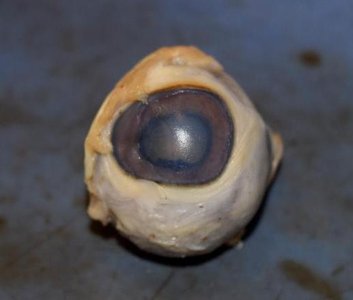
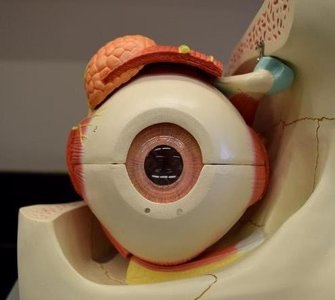
The external structures of the eye protect and support the function of vision. Key components include:
Sclera: The tough, white outer layer of the eyeball that provides structure and protection.
Cornea: The transparent, dome-shaped surface that covers the front of the eye and helps focus light.
Iris: The colored part of the eye, containing muscles that control the size of the pupil.
Lacrimal Glands: Glands located above the lateral end of each eye that produce tears for lubrication and protection.
Optic Nerve
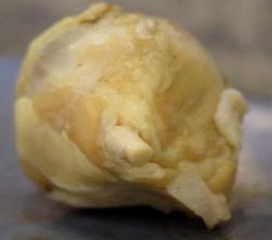
The optic nerve transmits visual information from the retina to the brain. It exits the posterior aspect of the eyeball and is essential for vision.
Internal Structures of the Eye
The internal structures of the eye are responsible for focusing light and converting it into neural signals. Major components include:
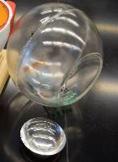
Lens: A transparent, flexible structure that focuses light onto the retina.
Vitreous Humor: The clear, gel-like substance that fills the space between the lens and the retina, maintaining the eye's shape.
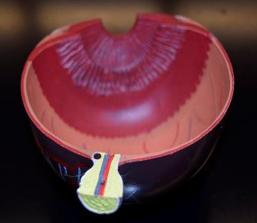
Internal Structures – Front
The anterior segment of the eye contains structures that regulate the amount of light entering the eye and help focus images:
Ciliary Body: Produces aqueous humor and contains muscles that control the shape of the lens for focusing (accommodation).
Pupillary Constrictor Muscle: Contracts to decrease pupil size in bright light (parasympathetic response).
Pupillary Dilator Muscle: Contracts to increase pupil size in dim light (sympathetic response).
Pupil: The opening in the center of the iris through which light enters the eye.
Internal Structures – Back
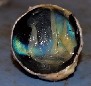
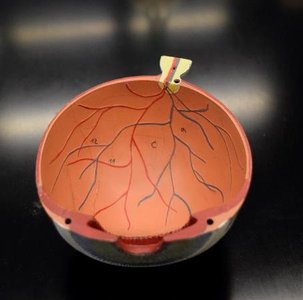
The posterior segment of the eye contains layers and structures essential for photoreception and signal transmission:
Choroid: Vascular layer that supplies nutrients and oxygen to the retina.
Retina: The innermost layer containing photoreceptor cells (rods and cones) that detect light and initiate neural signals.
Tapetum Lucidum: A reflective layer found in some animals (not humans) that enhances night vision.
Optic Disc: The point where the optic nerve exits the eye; also known as the "blind spot" because it lacks photoreceptors.
External Structures – Eye Muscles
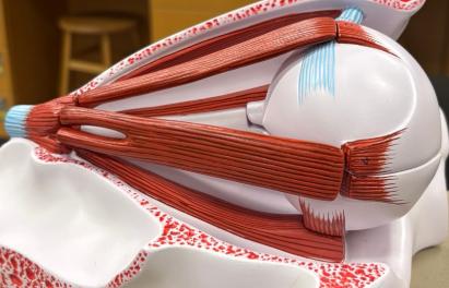
The movement of the eyeball is controlled by six extraocular muscles:
Lateral Rectus: Moves the eye laterally (outward).
Medial Rectus: Moves the eye medially (inward).
Superior Rectus: Elevates the eye and turns it medially.
Inferior Rectus: Depresses the eye and turns it medially.
Superior Oblique: Depresses the eye and turns it laterally.
Inferior Oblique: Elevates the eye and turns it laterally.
Ear
External Ear
The external ear collects sound waves and directs them toward the middle ear. Its main structures are:
Auricle (Pinna): The visible part of the ear that collects and directs sound waves into the ear canal.
External Acoustic Meatus (Ear Canal): The canal that conducts sound waves from the auricle to the tympanic membrane.
Tympanic Membrane (Eardrum): Vibrates in response to sound waves, transmitting vibrations to the middle ear.
Auditory Tube (Eustachian Tube): Connects the middle ear to the nasopharynx and equalizes air pressure.
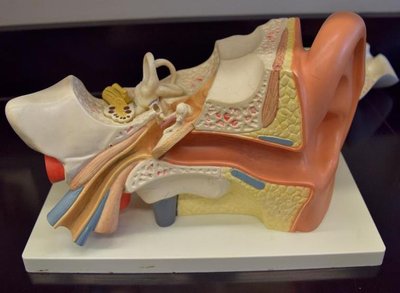
Inner Ear
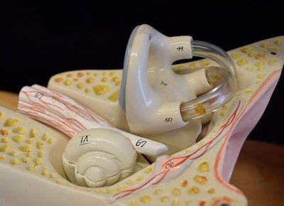
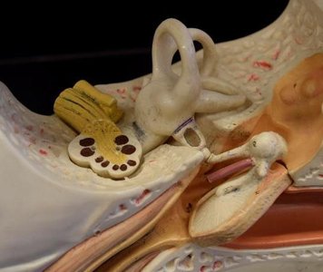
The inner ear contains structures responsible for hearing and balance:
Cochlea: A spiral-shaped organ that converts sound vibrations into neural signals for hearing.
Semicircular Canals: Three looped structures involved in detecting rotational movements and maintaining balance.
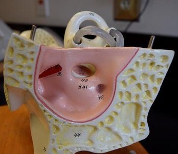
Vestibule: The central part of the bony labyrinth, involved in detecting linear acceleration and head position.
Cranial Nerve VIII (Vestibulocochlear Nerve): Transmits auditory and balance information from the inner ear to the brain.
Malleus, Incus, Stapes: The three auditory ossicles that transmit sound vibrations from the tympanic membrane to the inner ear.
Oval Window: The membrane-covered opening that leads from the middle ear to the vestibule of the inner ear.
Round Window: Another membrane-covered opening that allows for movement of fluid within the cochlea.
Summary Table: Key Structures of the Eye and Ear
Structure | Location | Function |
|---|---|---|
Sclera | Eye (outer layer) | Protection, shape maintenance |
Cornea | Eye (front) | Light refraction, protection |
Iris | Eye (front) | Controls pupil size |
Lens | Eye (internal) | Focuses light on retina |
Retina | Eye (internal) | Photoreception |
Optic Nerve | Eye (posterior) | Transmits visual signals |
Auricle | Ear (external) | Collects sound |
Tympanic Membrane | Ear (external/middle) | Transmits sound vibrations |
Cochlea | Ear (inner) | Hearing |
Semicircular Canals | Ear (inner) | Balance |
Additional info: The tapetum lucidum is present in many nocturnal animals to enhance vision in low light but is absent in humans. The pupillary muscles are controlled by the autonomic nervous system, with the constrictor muscle under parasympathetic control and the dilator muscle under sympathetic control.
