 뒤로
뒤로The Tissue Level of Organization: Histology and the Four Primary Tissue Types
스터디 가이드 - 스마트 노트
 자료에 맞춘 맞춤형 노트, 핵심 정의, 예시, 맥락을 확장해 제공합니다.
Histology and the Tissue Level of Organization
Introduction to Histology
Histology is the scientific study of tissues, focusing on their structure and function. Tissues are organized groups of cells and cell products that perform specific, limited functions. Understanding tissues is essential for comprehending how organs and organ systems operate within the human body.
Histology: The study of tissues, their structure, and function.
Tissues represent the next level of biological organization, situated between cells and organs.
There are four primary tissue types: epithelial, connective, muscle, and neural.
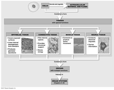
The Four Primary Tissue Types
Epithelial Tissue
Epithelial tissue covers body surfaces, lines internal cavities and passageways, and forms certain glands. It serves as a protective barrier and is involved in absorption, secretion, and sensation.
Locations: Epidermis of the skin, lining of the mouth, trachea, small intestine, blood vessels, heart chambers, and urinary passageways.
Characteristics:
Cellularity: Composed almost entirely of tightly packed cells with minimal extracellular material.
Polarity: Has distinct apical (top) and basal (bottom) surfaces.
Attachment: Basal surface attached to a basement membrane.
Avascularity: Lacks direct blood supply; nutrients diffuse from underlying tissues.
Regeneration: High capacity for renewal via stem cells, especially near the basal lamina.
Functions:
Physical protection
Control of permeability
Sensation
Production of specialized secretions (glandular epithelium)
Intercellular Connections
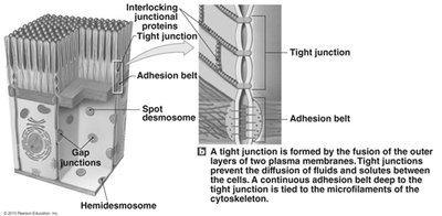
Cell junctions in epithelial tissue form bonds between cells or with extracellular material, contributing to tissue integrity and function.
Tight (occluding) junctions: Prevent passage of substances between cells.
Gap junctions: Allow communication between adjacent cells.
Desmosomes (macula adherens): Provide mechanical strength by anchoring cells together.
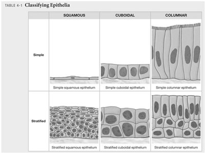
Classification of Epithelia
Epithelia are classified based on the number of cell layers and the shape of the cells at the apical surface.
By layers:
Simple epithelium: Single layer of cells
Stratified epithelium: Multiple layers of cells
By shape:
Squamous: Thin and flat
Cuboidal: Cube-shaped
Columnar: Tall and rectangular
Glandular Epithelia
Glandular epithelia form glands that produce secretions. Glands are classified as endocrine (secreting hormones into the blood) or exocrine (secreting onto surfaces through ducts).
Endocrine glands: Release hormones into interstitial fluid; ductless.
Exocrine glands: Release secretions onto epithelial surfaces via ducts.
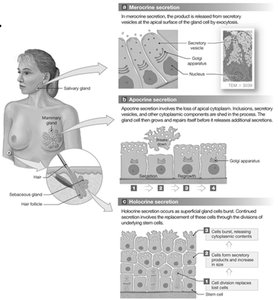
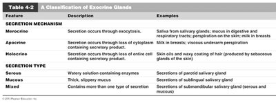
Mechanisms of Secretion
Merocrine secretion: Product released by exocytosis (e.g., sweat glands).
Apocrine secretion: Product released by shedding cytoplasm (e.g., mammary glands).
Holocrine secretion: Product released by cell bursting, destroying the cell (e.g., sebaceous glands).
Types of Exocrine Secretions
Serous glands: Watery secretions containing enzymes.
Mucous glands: Secrete mucins, forming thick, sticky mucus.
Mixed exocrine glands: Contain both serous and mucous secretions.
Feature | Description | Examples |
|---|---|---|
Merocrine | Secretion occurs through exocytosis | Saliva from salivary glands; sweat |
Apocrine | Secretion occurs through loss of cytoplasm | Milk in breasts; underarm perspiration |
Holocrine | Secretion occurs through loss of entire cell | Skin oils; sebaceous glands |
Serous | Watery solution containing enzymes | Parotid salivary gland |
Mucous | Thick, slippery mucus | Submandibular salivary gland |
Mixed | Contains more than one type of secretion | Submandibular salivary gland (serous and mucous) |
Connective Tissue
Connective tissue provides structural support, protection, and insulation for other tissues and organs. It is characterized by cells embedded in an extensive extracellular matrix composed of protein fibers and ground substance.
Main characteristics:
Specialized cells (e.g., fibroblasts, adipocytes, macrophages, mast cells)
Extracellular protein fibers (collagen, elastic, reticular)
Ground substance (fluid or semi-solid matrix)
Functions:
Support and protection (bone, cartilage)
Transport of materials (blood, lymph)
Energy storage (adipose tissue)
Defense (immune cells, antibodies)
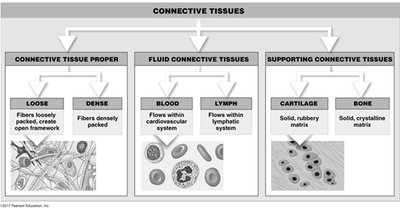
Types of Connective Tissue
Connective tissue proper: Loose (more ground substance, less fibers; e.g., adipose) and dense (more fibers, less ground substance; e.g., tendons)
Fluid connective tissue: Blood and lymph
Supporting connective tissue: Cartilage (gel-like matrix) and bone (calcified matrix)
Muscle Tissue
Muscle tissue is specialized for contraction, enabling movement of the body and its parts. It also generates heat as a byproduct of contraction. The two main contractile proteins are actin and myosin.
Skeletal muscle: Voluntary, striated, responsible for body movement.
Cardiac muscle: Involuntary, striated, found only in the heart.
Smooth muscle: Involuntary, non-striated, found in walls of hollow organs (e.g., blood vessels, digestive tract).
Neural Tissue
Neural tissue, also known as nervous tissue, is specialized for the conduction of electrical impulses. It rapidly senses changes in the internal and external environment and coordinates responses. Neural tissue is concentrated in the central nervous system (brain and spinal cord).
Neurons: Nerve cells that transmit electrical signals.
Neuroglia: Supporting cells that provide nutrients, repair, and protection for neurons.
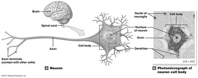
Structure of a Neuron
Cell body: Contains the nucleus and organelles.
Dendrites: Receive incoming signals.
Axon: Conducts outgoing electrical impulses to other cells.
Membranes
Types of Membranes
Membranes are physical barriers composed of epithelial and connective tissues. They line or cover portions of the body and are classified into four types:
Mucous membranes (mucosae): Line passageways with external connections (digestive, respiratory, urinary, reproductive tracts); contain goblet cells that secrete mucus.
Serous membranes: Line cavities not open to the outside; secrete serous fluid to reduce friction (e.g., pleura, pericardium, peritoneum).
Cutaneous membrane: The skin; thick, waterproof, and dry.
Synovial membranes: Line joint cavities; produce synovial fluid for lubrication; lack a true epithelium.
Tissue Injuries, Repair, and Aging
Inflammation and Regeneration
Tissues respond to injury through inflammation and regeneration to restore homeostasis.
Inflammation: The initial response to injury, characterized by swelling, redness, heat, and pain. It may be triggered by trauma or infection.
Regeneration: Replacement of damaged tissue with new cells.
Aging and Tissue Structure
With age, tissue repair slows, and the risk of cancer increases due to decreased metabolism, hormonal changes, and reduced physical activity. Environmental factors and genetics also contribute to cancer risk.
Key Terms and Cell Types
Fibroblasts: Produce fibers in connective tissue.
Osteocytes: Mature bone cells.
Adipocytes: Fat storage cells.
Chondrocytes: Cartilage cells.
Erythrocytes: Red blood cells; transport oxygen.
Thrombocytes: Platelets; involved in blood clotting.
Leukocytes: White blood cells; immune defense.
Goblet cells: Secrete mucus in mucous membranes.
Neuroglia: Support neurons.
Neuron: Conducts electrical impulses.
Summary Table: Classification of Epithelia
Squamous | Cuboidal | Columnar | |
|---|---|---|---|
Simple | Simple squamous epithelium | Simple cuboidal epithelium | Simple columnar epithelium |
Stratified | Stratified squamous epithelium | Stratified cuboidal epithelium | Stratified columnar epithelium |
Additional info: For further study, refer to textbook chapters on tissue structure, cell types, and the mechanisms of tissue repair and aging.
