 뒤로
뒤로The Tissue Level of Organization: Structure, Function, and Classification
스터디 가이드 - 스마트 노트
 자료에 맞춘 맞춤형 노트, 핵심 정의, 예시, 맥락을 확장해 제공합니다.
The Tissue Level of Organization
Introduction to Tissues
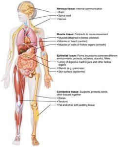
Tissues are groups of cells with a common embryonic origin that work together to perform specialized functions. Understanding tissue types is fundamental for grasping how cells organize into tissues, tissues into organs, and how tissue damage can lead to medical concerns.
Four Basic Tissue Types:
Epithelial Tissue: Covers body surfaces and lines cavities.
Connective Tissue: Supports, binds, and protects other tissues.
Muscle Tissue: Responsible for movement.
Nervous Tissue: Regulates and controls body functions.
Microscopy in Tissue Study
Techniques for Observing Tissues
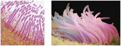
Microscopy is essential for studying tissue structure. Light microscopy uses colored dyes, while electron microscopy uses heavy metal coatings to visualize fine details.
Cell Junctions in Tissues
Types of Cell Junctions
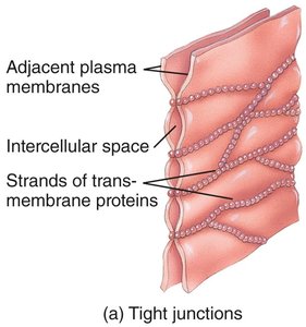
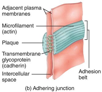
Cells in tissues are often bound together by specialized junctions, which help maintain tissue integrity and function.
Tight Junctions: Form impermeable barriers to prevent leakage between cells.
Adherens Junctions: Link cytoskeletal components for strong mechanical attachments.
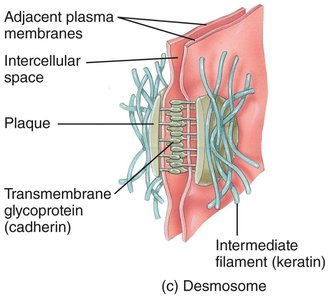
Desmosomes: Provide anchoring strength and allow tissues to resist tearing.
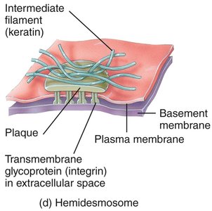
Hemidesmosomes: Attach cells to the basement membrane.
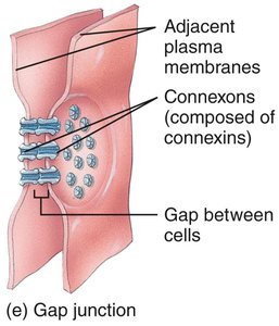
Gap Junctions: Allow communication and passage of small molecules between cells.
Epithelial Tissue
General Characteristics
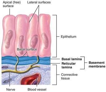
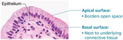
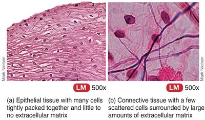
Epithelial tissue consists of sheets of closely packed cells with little extracellular matrix. It is avascular but innervated, and cells are attached to a basement membrane. Epithelial tissue is classified by cell shape and number of layers.
Main Functions: Protection, absorption, filtration, excretion, secretion, and sensory reception.
Polarity: Has apical (free) and basal (attached) surfaces.
Classification of Epithelia
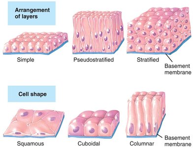
Epithelia are classified by the number of layers and cell shape:
Simple: One layer of cells.
Stratified: Two or more layers.
Pseudostratified: Appears multi-layered but all cells touch the basement membrane.
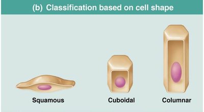
Squamous: Flat and scale-like.
Cuboidal: Cube-shaped.
Columnar: Tall and column-like.
Simple Epithelia
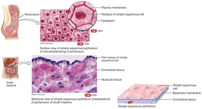
Simple Squamous Epithelium: Single layer of flat cells; found in air sacs of lungs, lining of blood vessels (endothelium), and serous membranes (mesothelium). Function: diffusion and filtration.
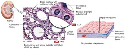
Simple Cuboidal Epithelium: Single layer of cube-shaped cells; found in kidney tubules and glandular ducts. Function: secretion and absorption.
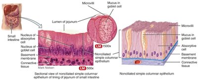
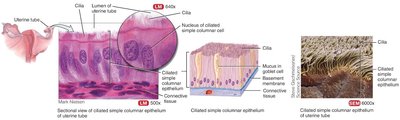
Simple Columnar Epithelium: Single layer of tall cells; may have microvilli or cilia. Nonciliated type lines digestive tract; ciliated type lines uterine tubes. Function: absorption and secretion.
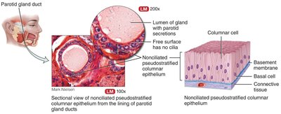
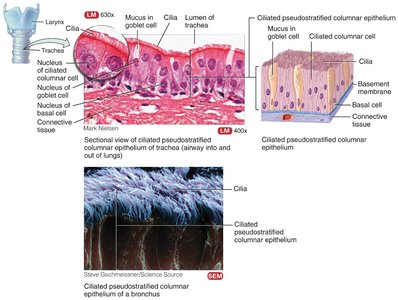
Pseudostratified Columnar Epithelium: Appears layered due to nuclei at different levels; all cells touch basement membrane. Nonciliated type lines ducts of large glands; ciliated type lines upper respiratory tract.
Stratified Epithelia
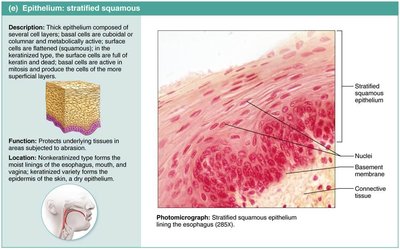
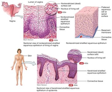
Stratified Squamous Epithelium: Multiple layers; protects against abrasion. Keratinized type forms skin; nonkeratinized type lines moist surfaces (mouth, esophagus, vagina).
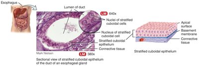
Stratified Cuboidal Epithelium: Usually two layers; found in ducts of sweat glands.
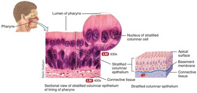
Stratified Columnar Epithelium: Limited distribution; found in male urethra and some glandular ducts.
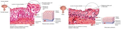
Transitional Epithelium: Found in urinary bladder; cells change shape to allow stretching.
Glandular Epithelium
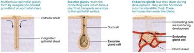
Endocrine Glands: Ductless; secrete hormones into blood (e.g., pituitary, thyroid).
Exocrine Glands: Secrete products into ducts (e.g., sweat, salivary glands).
Unicellular Glands: Single cells (e.g., goblet cells).
Multicellular Glands: Composed of many cells (e.g., salivary glands).
Connective Tissue
General Characteristics
Connective tissue is the most abundant and widely distributed tissue type. It consists of cells and an extracellular matrix (ECM) composed of fibers and ground substance. Functions include support, protection, insulation, energy storage, and transport.
Main Classes: Connective tissue proper, cartilage, bone, blood.
Major Fibers: Collagen (strength), elastic (stretch), reticular (support).
Cell Types: Fibroblasts, chondroblasts, osteoblasts, hematopoietic stem cells, adipocytes, white blood cells, mast cells, macrophages.
Types of Connective Tissue
Loose Connective Tissue: Areolar, adipose, reticular.
Dense Connective Tissue: Dense regular, dense irregular, elastic.
Cartilage: Hyaline, fibrocartilage, elastic.
Bone: Compact and spongy bone.
Blood: Fluid connective tissue with plasma and formed elements.
Cartilage
Hyaline Cartilage: Most abundant; found at ends of long bones, nose, trachea.
Fibrocartilage: Strongest; found in intervertebral discs, knee menisci.
Elastic Cartilage: Flexible; found in ear and epiglottis.
Bone (Osseous Tissue)
Structure: Osteons in compact bone; trabeculae in spongy bone.
Function: Support, protection, storage, blood cell formation.
Blood
Components: Plasma, red blood cells, white blood cells, platelets.
Function: Transport of gases, nutrients, wastes, immune responses, clotting.
Muscle Tissue
Types of Muscle Tissue
Skeletal Muscle: Voluntary, striated, multinucleated; attached to bones for movement.
Cardiac Muscle: Involuntary, striated, single nucleus, intercalated discs; found in heart.
Smooth Muscle: Involuntary, non-striated, single nucleus; found in walls of hollow organs.
Nervous Tissue
Structure and Function
Nervous tissue is specialized for communication and control. It consists of neurons (which generate and conduct impulses) and neuroglia (supporting cells).
Neurons: Cell body, dendrites, axon; transmit sensory and motor information.
Neuroglia: Support, insulate, and protect neurons.
Membranes
Types of Membranes
Epithelial Membranes: Mucous, serous, and cutaneous membranes.
Synovial Membranes: Line joint cavities; produce synovial fluid.
Tissue Repair
Regeneration and Fibrosis
Tissue repair replaces damaged or dead cells. Epithelial and some connective tissues regenerate well; muscle and nervous tissues have limited repair capacity. Fibrosis results in scar tissue formation.
Embryonic Connective Tissue
Types
Mesenchyme: Gives rise to all other connective tissues.
Mucous Connective Tissue: Found in umbilical cord.
