 뒤로
뒤로Tissue: The Living Fabric – Epithelial Tissue Study Guide
스터디 가이드 - 스마트 노트
 자료에 맞춘 맞춤형 노트, 핵심 정의, 예시, 맥락을 확장해 제공합니다.
Tissue: The Living Fabric
Introduction to Tissues
Tissues are groups of cells with similar structure and function, working together to maintain homeostasis in the body. The study of tissues is known as histology. There are four primary tissue types: epithelial, connective, muscle, and nervous tissue.
Epithelial tissue: Covers surfaces and forms boundaries.
Connective tissue: Supports, protects, and binds other tissues.
Muscle tissue: Produces movement.
Nervous tissue: Controls and communicates.
Types of Primary Tissues
Epithelial tissue: Forms boundaries, protects, secretes, absorbs, and filters. Examples include the skin surface (epidermis) and lining of digestive tract organs.
Connective tissue: Supports and binds other tissues. Examples include bones, tendons, and fat.
Muscle tissue: Contracts to cause movement. Found in skeletal muscles, heart, and walls of hollow organs.
Nervous tissue: Internal communication. Found in the brain, spinal cord, and nerves.
Studying Human Tissue: Microscopy
Microscopy is essential for studying tissues. Tissue samples are preserved (fixed), sliced thinly (cut), and stained to enhance contrast for light or electron microscopy.
Epithelial Tissue
Overview and Functions
Epithelial tissue forms boundaries and is classified by location as covering/lining epithelia (external and internal surfaces) or glandular epithelia (secretory tissue in glands). Its functions include:
Protection
Absorption
Filtration
Excretion
Secretion
Sensory reception
Five Characteristics of Epithelial Tissues
Polarity: Cells have an apical (upper, free) surface and a basal (lower, attached) surface, each with distinct structure and function.
Specialized contacts: Cells fit closely together, forming continuous sheets with tight junctions and desmosomes.
Supported by connective tissues: The basement membrane (basal lamina + reticular lamina) reinforces the epithelial sheet.
Avascular but innervated: No blood vessels; nutrients diffuse from underlying connective tissue. Supplied by nerve fibers.
Regeneration: High capacity for regeneration, especially when exposed to friction or hostile environments.
Polarity in Epithelial Tissue
Apical surface: May be smooth, have microvilli (increase surface area, e.g., intestinal lining), or cilia (move substances, e.g., trachea).
Basal surface: Noncellular basal lamina acts as an adhesive sheet, selective filter, and scaffolding for cell migration.
Specialized Contacts
Tight junctions: Prevent leakage between cells.
Desmosomes: Provide mechanical strength.
Connective Tissue Support
Basement membrane: Combination of basal and reticular lamina; resists stretching and tearing, defines epithelial boundary.
Avascular but Innervated
Avascular: No direct blood supply; nutrients diffuse from underlying tissues.
Innervated: Contains nerve fibers for sensation.
Regeneration
High regenerative capacity: Cells divide to replace lost or damaged cells, especially in areas exposed to friction.
Classification of Epithelia
By Number of Cell Layers
Simple epithelia: Single layer of cells; functions in absorption, secretion, filtration.
Stratified epithelia: Two or more layers; functions in protection.
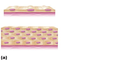
By Cell Shape
Squamous: Flattened, scale-like cells.
Cuboidal: Box-like cells with round nuclei.
Columnar: Tall, column-shaped cells with elongated nuclei.
Types of Epithelial Tissue
Simple Epithelia
Simple Squamous Epithelium
Single layer of flattened cells with disc-shaped nuclei and sparse cytoplasm. Functions in rapid diffusion and filtration.
Location: Kidney glomeruli, air sacs of lungs, lining of heart, blood vessels, lymphatic vessels, serosae.
Special types: Endothelium (lining of vessels and heart), Mesothelium (serous membranes).
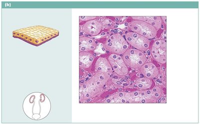
Simple Cuboidal Epithelium
Single layer of cubelike cells with large, spherical nuclei. Functions in secretion and absorption.
Location: Kidney tubules, ducts and secretory portions of small glands, ovary surface.
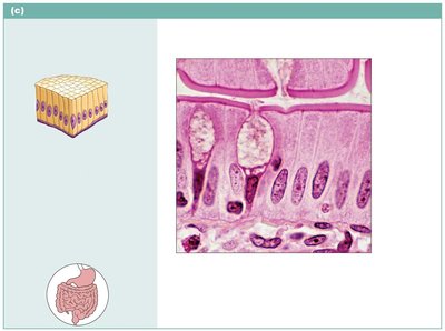
Simple Columnar Epithelium
Single layer of tall cells with round to oval nuclei; may have cilia and goblet cells. Functions in absorption and secretion of mucus, enzymes, and other substances.
Location: Nonciliated type lines most of digestive tract, gallbladder, excretory ducts; ciliated type lines small bronchi, uterine tubes, uterus.
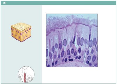
Pseudostratified Columnar Epithelium
Single layer of cells of varying heights; nuclei at different levels. Functions in secretion (especially mucus) and propulsion by ciliary action.
Location: Nonciliated type in male’s sperm-carrying ducts and large glands; ciliated type lines trachea and upper respiratory tract.
Stratified Epithelial Tissues
Stratified Squamous Epithelium
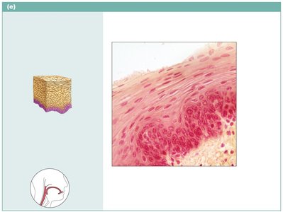
Thick membrane with several cell layers; basal cells are cuboidal/columnar, surface cells are squamous. Functions in protection against abrasion.
Location: Nonkeratinized type in esophagus, mouth, vagina; keratinized type forms epidermis of skin.
Stratified Cuboidal Epithelium
Rare; typically two cell layers thick. Found in some sweat and mammary glands.
Stratified Columnar Epithelium
Limited distribution; found in pharynx, male urethra, and some glandular ducts. Only apical layer is columnar.
Transitional Epithelium
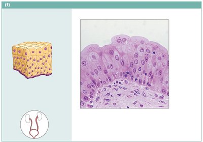
Resembles both stratified squamous and cuboidal; basal cells cuboidal/columnar, surface cells dome-shaped or squamous-like depending on stretch. Functions in stretching and distension of urinary organs.
Location: Lines ureters, bladder, and part of urethra.
Glandular Epithelia
Overview of Glands
A gland is one or more cells that makes and secretes an aqueous fluid called a secretion. Glands are classified by site of product release (endocrine or exocrine) and number of cells (unicellular or multicellular).
Endocrine Glands
Ductless glands: Secretions (hormones) released into blood or lymph, targeting specific organs.
Exocrine Glands
Secretions released onto body surfaces or into cavities: More numerous than endocrine glands; secrete products into ducts. Examples: mucous, sweat, oil, salivary glands.
Unicellular Exocrine Glands
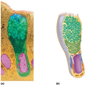
Mucous cells and goblet cells: Found in epithelial linings of intestinal and respiratory tracts; produce mucin, which forms mucus for protection and lubrication.
Multicellular Exocrine Glands
Composed of a duct and secretory unit; usually surrounded by supportive connective tissue.
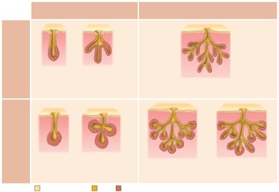
Classified by structure (simple or compound; tubular, alveolar, or tubuloalveolar) and type of secretion (merocrine, holocrine, apocrine).
Classification of Multicellular Glands
Structure | Example |
|---|---|
Simple tubular | Intestinal glands |
Simple branched tubular | Stomach (gastric) glands |
Compound tubular | Duodenal glands of small intestine |
Simple alveolar | No important example in humans |
Simple branched alveolar | Sebaceous (oil) glands |
Compound alveolar | Mammary glands |
Compound tubuloalveolar | Salivary glands |
Modes of Secretion
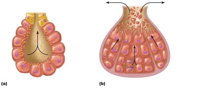
Merocrine: Secrete products by exocytosis as produced (e.g., sweat glands).
Holocrine: Accumulate products within, then rupture (e.g., sebaceous glands).
Apocrine: Accumulate products within, only apex ruptures (controversial in humans).
Summary Table: Epithelial Tissue Types
Type | Description | Function | Location |
|---|---|---|---|
Simple squamous | Single layer, flat cells | Diffusion, filtration | Lungs, vessels, serosae |
Simple cuboidal | Single layer, cube cells | Secretion, absorption | Kidney tubules, glands |
Simple columnar | Single layer, tall cells | Absorption, secretion | Digestive tract, bronchi |
Pseudostratified columnar | Single layer, varying heights | Secretion, propulsion | Trachea, upper respiratory |
Stratified squamous | Multiple layers, flat cells | Protection | Skin, mouth, esophagus |
Stratified cuboidal | Two layers, cube cells | Protection | Sweat, mammary glands |
Stratified columnar | Multiple layers, tall cells | Protection, secretion | Pharynx, urethra |
Transitional | Multiple layers, variable shape | Stretching | Bladder, ureters |
Key Terms
Histology: Study of tissues
Basal lamina: Adhesive sheet, selective filter
Basement membrane: Basal lamina + reticular lamina
Goblet cell: Unicellular gland producing mucus
Merocrine, holocrine, apocrine: Modes of gland secretion
Example
Simple squamous epithelium in the alveoli of the lungs allows for rapid gas exchange by diffusion, while stratified squamous epithelium in the skin provides protection against abrasion.
Additional info: The notes expand on brief points from the slides, providing full academic context, definitions, and examples for each tissue type and classification.
