 뒤로
뒤로Tissues and Cell Junctions: Structure and Function in Human Anatomy
스터디 가이드 - 스마트 노트
 자료에 맞춘 맞춤형 노트, 핵심 정의, 예시, 맥락을 확장해 제공합니다.
Tissues: Definition and Levels of Organization
Definition and Placement in Biological Hierarchy
Tissues are collections of specialized cells and cell products that perform specific functions. They represent a fundamental level of biological organization, falling between cells and organs. Histology is the scientific study of tissues. - Tissue: A group of similar cells working together to perform a specific function. - Levels of Organization: Atoms & molecules → Cells → Tissues → Organs → Organ systems → Organism. - Example: Cardiac muscle tissue forms the heart, which is part of the cardiovascular system. 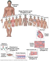
The Four General Tissue Types
There are four primary tissue types in the human body, each with distinct functions and locations. - Epithelial Tissue: Covers exposed surfaces and lines internal passageways. - Connective Tissue: Fills internal spaces, supports other tissues, transports materials, and stores energy. - Muscle Tissue: Specialized for contraction, found in skeletal muscle, heart muscle, and walls of hollow organs. - Nervous Tissue: Carries electrical signals throughout the body. 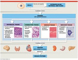
Epithelial Tissue: Structure and Function
Characteristics and Functions
Epithelial tissue includes both epithelia and glands. Epithelia are layers of cells covering internal or external surfaces, while glands produce fluid secretions. - Functions: Physical protection, control of permeability, sensation, and production of specialized secretions. - Characteristics: Polarity (apical and basal surfaces), cellularity (cell junctions), attachment (basement membrane), avascularity, and regeneration.
Specializations of Epithelial Cells
Epithelial cells exhibit specializations that enhance their function. - Apical Surface: Faces the environment or lumen; may have microvilli (increase absorption/secretion) or cilia (move fluids). - Basal Surface: Anchors the cell to underlying tissues. - Cell Junctions: Maintain tissue integrity and communication. 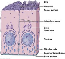
Identifying Epithelial Tissue in Microscopy
When examining slides, epithelial tissues are identified by their apical surface, which is exposed to the environment or a lumen. - Microscopy Tip: Scan for the apical surface at low magnification (4X) to locate epithelial tissue. 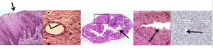
Maintenance and Repair of Epithelia
The integrity of epithelial tissue is maintained by intercellular connections, attachment to the basement membrane, and regenerative capacity. 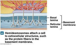
Cell Junctions: Types and Functions
Overview of Cell Junctions
Cell junctions are intercellular connections that form bonds between cells or between cells and extracellular material. The main types are gap junctions, tight junctions, and desmosomes.
Gap Junctions
Gap junctions allow rapid communication between cells by permitting the passage of ions and small molecules. - Structure: Cells are held together by interlocking transmembrane proteins called connexons. - Function: Coordinate contractions in heart muscle and other tissues. 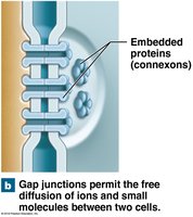
Tight Junctions
Tight junctions are formed by the fusion of the outer layers of two plasma membranes. - Function: Prevent passage of water and solutes, keeping enzymes, acids, and wastes in the lumen of the digestive tract. - Adhesion Belt: Attaches to the terminal web, reinforcing the tight junction. 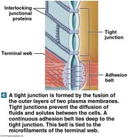
Desmosomes
Desmosomes are strong intercellular junctions that tie cells together and allow bending and twisting. - Spot Desmosomes: Tie adjacent cells together using cell adhesion molecules (CAMs) and proteoglycans. - Hemidesmosomes: Attach cells to the basement membrane, anchoring them to extracellular structures. 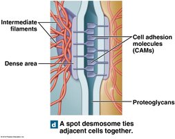
Summary Table: Types of Cell Junctions
Junction Type | Structure | Function | Location |
|---|---|---|---|
Gap Junction | Connexons (protein channels) | Rapid communication, passage of ions | Cardiac muscle, some epithelia |
Tight Junction | Interlocking junctional proteins, adhesion belt | Barrier to fluid/solute passage | Digestive tract, epithelia |
Spot Desmosome | CAMs, proteoglycans, intermediate filaments | Strong attachment, flexibility | Skin, heart muscle |
Hemidesmosome | Basal lamina, reticular lamina | Anchors cell to basement membrane | Basal surface of epithelia |
Conclusion
Understanding the structure and function of tissues and cell junctions is fundamental to the study of anatomy and physiology. These concepts provide the basis for comprehending how cells organize into tissues, how tissues form organs, and how organs work together in organ systems to maintain the health and function of the human body. Additional info: Academic context was added to clarify the hierarchy of organization, the functional significance of cell junctions, and the identification of epithelial tissues in microscopy.
