 뒤로
뒤로Tissues and Early Embryology: Structure and Function of Human Tissues
스터디 가이드 - 스마트 노트
 자료에 맞춘 맞춤형 노트, 핵심 정의, 예시, 맥락을 확장해 제공합니다.
Chapter 3: Tissues and Early Embryology
Introduction to Tissues
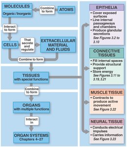
Tissues are groups of cells with similar structure and function, forming the foundation for organs and organ systems in the human body. Understanding tissue types is essential for comprehending how the body is organized and how it functions at a microscopic level.
Epithelial Tissue
General Characteristics and Functions
Epithelial tissue consists of sheets of cells that cover exposed surfaces and line internal cavities and passageways. These tissues serve as protective barriers and are involved in absorption, secretion, and sensation.
Physical protection: Shields underlying tissues from mechanical and chemical stress.
Control permeability: Regulates the movement of substances into and out of the body.
Provide sensation: Contains sensory nerve endings for detecting changes in the environment.
Produce secretions: Forms glands that secrete substances such as enzymes, hormones, and mucus.
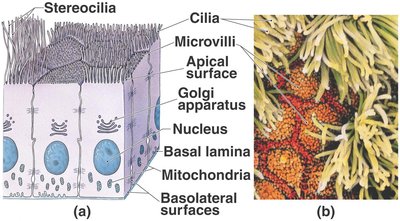
Specializations of epithelial cells include:
Microvilli: Increase surface area for absorption.
Stereocilia: Long microvilli, found in the male reproductive tract and inner ear.
Ciliated epithelium: Moves substances across the epithelial surface.
Maintaining Epithelial Integrity
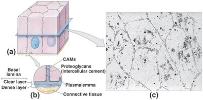
Epithelial tissues maintain their integrity through specialized intercellular connections and attachment to the basal lamina. The basal lamina consists of two layers: the lamina lucida (clear layer) and the lamina densa (dense layer). Epithelial cells are capable of self-renewal through mitosis.
Classification of Epithelia
Epithelia are classified based on the number of cell layers and the shape of the cells:
Simple epithelium: One layer of cells.
Stratified epithelium: Multiple layers of cells.
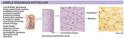
Squamous: Thin, flat cells.
Cuboidal: Cells as tall as they are wide.
Columnar: Cells taller than they are wide.
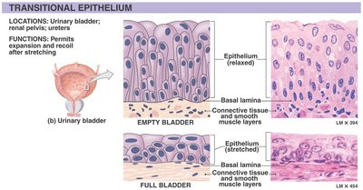
Transitional: Cells that change shape (e.g., in the urinary bladder).
Examples of Epithelial Types
Type | Location | Function |
|---|---|---|
Simple Squamous | Alveoli of lungs, lining of heart and blood vessels | Reduces friction, controls vessel permeability, absorption, secretion |
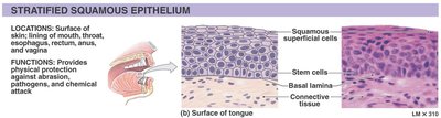
Stratified Squamous | Surface of skin, lining of mouth, throat, esophagus | Physical protection against abrasion, pathogens, chemical attack |
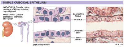
Simple Cuboidal | Glands, ducts, kidney tubules | Limited protection, secretion, absorption |
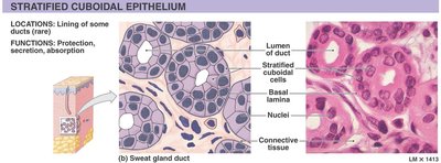
Stratified Cuboidal | Some ducts (rare) | Protection, secretion, absorption |
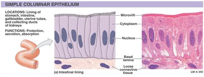
Simple Columnar | Lining of stomach, intestine, gallbladder | Protection, secretion, absorption |
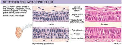
Stratified Columnar | Small areas of pharynx, anus, mammary gland ducts | Protection |
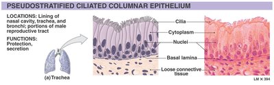
Pseudostratified Ciliated Columnar | Lining of nasal cavity, trachea, bronchi | Protection, secretion |
Transitional | Urinary bladder, renal pelvis, ureters | Permits expansion and recoil after stretching |
Glandular Epithelia
Glandular epithelia are specialized for secretion. Glands can be classified by the nature of their secretions and their structure:
Serous glands: Secrete watery fluids rich in enzymes.
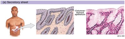
Mucous glands: Secrete mucins that absorb water to form mucus.
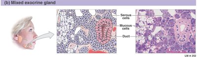
Mixed exocrine glands: Contain both serous and mucous cells.
Structural Classification of Glands
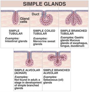
Simple Glands | Examples |
|---|---|
Simple tubular | Intestinal glands |
Simple coiled tubular | Merocrine sweat glands |
Simple branched tubular | Gastric glands, mucous glands of esophagus |
Simple alveolar (acinar) | Not found in adults |
Simple branched alveolar | Sebaceous (oil) glands |
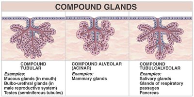
Compound Glands | Examples |
|---|---|
Compound tubular | Mucous glands (mouth), testes |
Compound alveolar (acinar) | Mammary glands |
Compound tubuloalveolar | Salivary glands, pancreas |
Mechanisms of Glandular Secretion
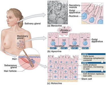
Merocrine secretion: Product released from secretory vesicles by exocytosis (e.g., salivary glands).
Apocrine secretion: Involves the loss of cytoplasm as well as the secretory product (e.g., mammary glands).
Holocrine secretion: Entire cell becomes packed with secretory products and then bursts (e.g., sebaceous glands).
Connective Tissues
General Structure and Functions
Connective tissues are diverse and provide structural and metabolic support for other tissues and organs. All connective tissues share three main components: specialized cells, extracellular protein fibers, and ground substance. The combination of fibers and ground substance forms the matrix.
Establish structural framework
Transport fluids and dissolved materials
Protect organs
Support, surround, and connect other tissues
Store energy
Defend the body from microorganisms
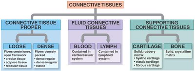
Classification of Connective Tissues
Type | Subtypes | Main Features |
|---|---|---|
Connective Tissue Proper | Loose, Dense | Loose: open framework; Dense: tightly packed fibers |
Fluid Connective Tissues | Blood, Lymph | Cells suspended in a watery matrix |
Supporting Connective Tissues | Cartilage, Bone | Dense matrix of fibers and ground substance |
Connective Tissue Proper: Cells and Fibers
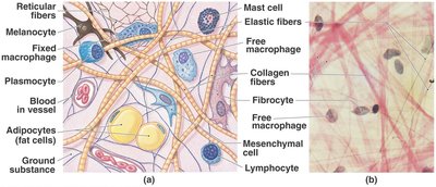
Connective tissue proper contains a variety of cell types and fibers:
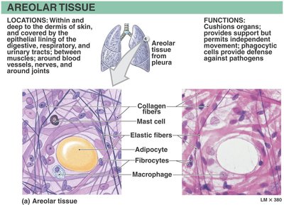
Collagen fibers: Strong, flexible, and the most common fiber type.
Reticular fibers: Form a supportive network.
Elastic fibers: Contain elastin, allowing tissues to stretch and recoil.
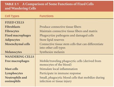
Cell Types in Connective Tissue Proper
Cell Type | Function |
|---|---|
Fibroblasts | Produce connective tissue fibers |
Fibrocytes | Maintain fibers and matrix |
Adipocytes | Store lipids |
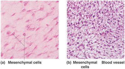
Mesenchymal cells | Stem cells for repair |
Macrophages | Phagocytize pathogens and debris |
Mast cells | Stimulate inflammation |
Lymphocytes | Participate in immune response |
Melanocytes | Synthesize melanin |
Loose and Dense Connective Tissue
Loose connective tissue: Areolar, adipose, and reticular tissues; provide support and cushioning.
Dense connective tissue: Dense regular (tendons, ligaments), dense irregular (dermis, organ capsules), and elastic tissue.
Fluid Connective Tissues
Blood and lymph are fluid connective tissues. Blood contains a matrix called plasma and formed elements (red cells, white cells, platelets). Lymph is formed from interstitial fluid and returns to the bloodstream via lymphatic vessels.
Supporting Connective Tissues
Supporting connective tissues include cartilage (hyaline, elastic, fibrous) and bone. These tissues provide structural support and protection for the body.
Membranes
Types of Membranes
Mucous membranes: Line passageways open to the exterior; moist surfaces.
Serous membranes: Line ventral body cavities; secrete serous fluid to reduce friction.
Cutaneous membrane: The skin; thick, dry, and water-resistant.
Synovial membranes: Line joint cavities; produce synovial fluid for lubrication.
Muscle and Neural Tissue
Muscle Tissue
Muscle tissue is specialized for contraction and includes three types:
Skeletal muscle: Voluntary movement, striated appearance.
Cardiac muscle: Involuntary, found in the heart, striated with intercalated discs.
Smooth muscle: Involuntary, found in walls of hollow organs, non-striated.
Neural Tissue
Neural tissue is specialized for the conduction of electrical impulses. It consists of two main cell types:
Neurons: Transmit electrical signals.
Neuroglia: Support, protect, and nourish neurons.
Tissues, Nutrition, and Aging
Effects of Aging on Tissues
As the body ages, tissue repair and maintenance become less efficient. Epithelia thin, connective tissues become fragile, and some tissues (such as cardiac muscle and neural tissue) cannot regenerate, leading to cumulative damage and potential health issues.
Embryology Summary
Early embryology explains the origin of tissues from the three primary germ layers: ectoderm, mesoderm, and endoderm. These layers give rise to all tissues and organs in the body.
Additional info: For further study, refer to histological slides and diagrams to reinforce the microscopic appearance and classification of tissues.
