 뒤로
뒤로Tissues: Structure, Classification, and Function in Anatomy & Physiology
스터디 가이드 - 스마트 노트
 자료에 맞춘 맞춤형 노트, 핵심 정의, 예시, 맥락을 확장해 제공합니다.
Review of Biological Levels of Organization
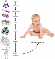
Hierarchy of Structural Organization
The human body is organized into a hierarchy of structural levels, each building upon the previous. Understanding these levels is essential for studying anatomy and physiology, as each level contributes to the function of the organism as a whole.
Chemical Level: Atoms combine to form molecules, which are the building blocks of cells.
Cellular Level: Molecules form organelles, which make up cells—the basic units of life.
Tissue Level: Similar cells and their extracellular matrix combine to form tissues with specialized functions.
Organ Level: Different tissues combine to form organs, each with specific functions.
Organ System Level: Organs interact in organ systems to perform complex functions.
Organism Level: All organ systems work together to maintain the life of the whole organism.
Tissues of the Body
Overview of Tissue Types
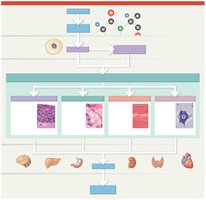
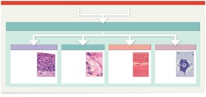
Tissues are groups of similar cells that perform a common function. The four primary tissue types in the human body are:
Epithelial Tissue: Covers exposed surfaces, lines internal passageways, and forms glands.
Connective Tissue: Fills internal spaces, provides structural support, stores energy, and defends the body.
Muscle Tissue: Contracts to produce movement.
Neural Tissue: Conducts electrical impulses and processes information.
Specialization and Organization of Tissues
Each tissue type is specialized for particular functions and is composed of cells and extracellular material. Tissues combine to form organs, which then interact within organ systems.
Epithelial Tissue
Functions of Epithelial Tissue
Epithelial tissue serves several key functions in the body:
Physical Protection: Shields underlying tissues from abrasion, dehydration, and destruction.
Control of Permeability: Regulates the movement of substances into and out of the body or organs.
Provides Sensation: Contains sensory receptors for detecting changes in the environment.
Produces Secretions: Forms glands that secrete substances such as enzymes, hormones, and mucus.
Characteristics of Epithelial Tissue
Cellularity: Composed almost entirely of tightly packed cells with minimal extracellular material.
Polarity: Has an apical (exposed) surface and a basal (attached) surface.
Attachment: The basal surface is attached to a basal lamina, separating it from underlying connective tissue.
Avascularity: Lacks blood vessels; nutrients diffuse from underlying tissues.
Arranged in Sheets: Cells are organized in one or more layers.
Regeneration: High capacity for cell division and repair.
Polarity and Surface Specializations
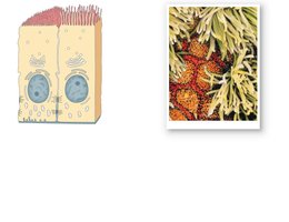
Epithelial cells exhibit polarity, with distinct apical and basal surfaces. The apical surface may have specializations such as microvilli (increase surface area for absorption) or cilia (move substances across the surface).
Attachment to Basal Lamina
The basal surface of epithelial cells is anchored to a basal lamina, which separates the epithelium from underlying connective tissue. This attachment provides structural support and regulates cell behavior.
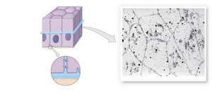
Cellularity and Intercellular Connections
Epithelial cells are tightly joined by specialized junctions, including tight junctions, adherens junctions, desmosomes, and gap junctions. These connections maintain tissue integrity and regulate communication between cells.
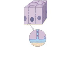
Specializations of Epithelial Cells
Microvilli, Stereocilia, and Cilia
Microvilli: Increase surface area for absorption (e.g., in the intestines).
Stereocilia: Long, non-motile microvilli found in the inner ear and male reproductive tract.
Cilia: Motile projections that move substances across the epithelial surface (e.g., in the respiratory tract).
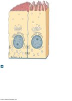
Classification of Epithelia
By Number of Layers and Cell Shape
Epithelia are classified based on the number of cell layers and the shape of the cells at the apical surface:
Simple Epithelium: Single layer of cells; all cells touch the basal lamina.
Stratified Epithelium: Multiple layers; only the deepest layer touches the basal lamina.
Cell Shapes: Squamous (flat), cuboidal (cube-shaped), columnar (tall and column-like).
Connective Tissue
Functions of Connective Tissue
Connective tissues provide a variety of functions essential for the structure and function of the body:
Structural Framework: Supports and anchors organs and tissues.
Transport: Blood and lymph transport nutrients, gases, and wastes.
Protection: Cushions and protects organs.
Support and Connection: Connects and supports other tissues.
Energy Storage: Stores energy in the form of fat.
Defense: Defends against pathogens and repairs tissue damage.
Classification of Connective Tissues
Connective tissues are classified into three main categories:
Connective Tissue Proper: Includes loose (areolar, adipose, reticular) and dense (regular, irregular, elastic) connective tissues.
Fluid Connective Tissue: Includes blood and lymph.
Supporting Connective Tissue: Includes cartilage and bone.
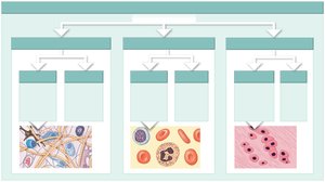
Cells and Fibers of Connective Tissue Proper
Connective tissue proper contains various cell types (fibroblasts, adipocytes, macrophages, etc.) and fibers (collagen, elastic, reticular) embedded in ground substance.
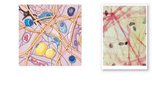
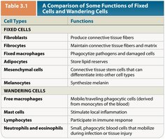
Comparison of Fixed and Wandering Cells
Connective tissue contains both fixed cells (e.g., fibroblasts, adipocytes) and wandering cells (e.g., macrophages, mast cells) that contribute to tissue maintenance, defense, and repair.
Cell Types | Functions |
|---|---|
Fibroblasts | Produce connective tissue fibers |
Fibrocytes | Maintain connective tissue fibers and matrix |
Fixed macrophages | Phagocytize pathogens and damaged cells |
Adipocytes | Store lipid reserves |
Mesenchymal cells | Connective tissue stem cells that differentiate into other cell types |
Melanocytes | Synthesize melanin |
Free macrophages | Mobile phagocytic cells |
Mast cells | Stimulate local inflammation |
Lymphocytes | Participate in immune response |
Neutrophils and eosinophils | Mobilize during infection or tissue injury |
Summary of Early Embryology: The Formation of Tissues
Origin of Tissues from Germ Layers
During embryonic development, three primary germ layers form: ectoderm, mesoderm, and endoderm. These layers give rise to all tissues and organs in the body:
Ectoderm: Forms neural tissue, epithelia, and glands.
Mesoderm: Forms connective tissues and muscle tissue.
Endoderm: Contributes to the formation of internal organs and their linings.
All three germ layers interact to produce functional organs and organ systems.
