Ch. 13 The Peripheral Nervous System and Reflex Activity
Marieb - Human Anatomy & Physiology 7th Edition
Marieb, Hoehn7th EditionHuman Anatomy & PhysiologyISBN: 9780805359091당신이 사용하는 게 아니라요?교과서 변경
13장, 문제 7

The portion of the fibrous layer that is white and opaque is the
a. Choroid
b. Cornea
c. Retina
d. Sclera

검증된 단계별 안내
1
Understand the structure of the fibrous layer of the eye, which consists mainly of two parts: the cornea and the sclera.
Recall that the cornea is the transparent, clear part of the fibrous layer that allows light to enter the eye.
Recognize that the sclera is the white, opaque portion of the fibrous layer, providing protection and structural support to the eye.
Eliminate options that are not part of the fibrous layer: the choroid is part of the vascular layer, and the retina is the innermost neural layer.
Conclude that the white and opaque portion of the fibrous layer is the sclera.

비슷한 문제에 대한 검증된 영상 답변:

이 영상 해법은 위 문제에 도움이 된다고 튜터들이 추천한 것입니다.
영상 길이:
2m

주요 개념

질문에 올바르게 답하기 위해 반드시 이해해야 하는 핵심 개념들은 다음과 같습니다.

Fibrous Layer of the Eye

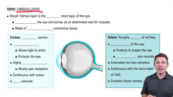
The fibrous layer is the outermost layer of the eyeball, providing structural support and protection. It consists mainly of two parts: the sclera and the cornea. Understanding this layer is essential to identify which part is white and opaque.
추천 영상:
5:59
Fibrous Layer

Sclera

The sclera is the white, opaque portion of the fibrous layer. It covers most of the eyeball and maintains the shape of the eye while protecting internal components. Its opacity distinguishes it from the transparent cornea.
추천 영상:
5:59
Fibrous Layer

Cornea

The cornea is the transparent, dome-shaped front part of the fibrous layer that allows light to enter the eye. Unlike the sclera, it is clear and plays a key role in focusing vision. Recognizing its transparency helps differentiate it from the sclera.
추천 영상:
5:59
Fibrous Layer
관련 실천
교과서 질문

Match the names of the cranial nerves in column B to the appropriate description in column A.

Column A 

_______    (1) Causes pupillary constriction 

_______    (2) The major sensory nerve of the face

_______    (3) Serves the sternocleido-mastoid and trapezius muscles 

_______    (4) Purely sensory (two nerves) 

_______    (5) Serves the tongue muscles 

_______    (6) Allows you to chew your food 

_______    (7) Impaired in Bell's palsy

_______    (8) Helps regulate heart activity

_______    (9) Helps you hear and maintain your balance 

_______   (10) Contain parasympathetic motor fibers (four nerves)

Column B

a. Abducens

b. Accessory

c. Facial

d. Glossopharyngeal

e. Hypoglossal

f. Oculomotor

g. Olfactory

h. Optic

i. Trigeminal

j. Trochlear

k. Vagus

l. Vestibulocochlear

1053
views
교과서 질문

The connective tissue sheath that surrounds a fascicle of nerve fibers is the

a. Epineurium

b. Endoneurium

c. Perineurium

d. Epimysium

1461
views
교과서 질문

Accessory glands that produce an oily secretion are the

a. Conjunctiva

b. Lacrimal glands

c. Tarsal glands

159
views
교과서 질문

Which sequence best describes a normal route for the flow of tears from the eyes into the nasal cavity?

a. Lacrimal canaliculi, lacrimal sacs, nasolacrimal ducts

b. Lacrimal ducts, lacrimal canaliculi, nasolacrimal ducts

c. Nasolacrimal ducts, lacrimal canaliculi, lacrimal sacs

1042
views
교과서 질문

Of the neurons in the retina, the axons of which of these form the optic nerve?

a. Bipolar cells

b. Ganglion cells

c. Cone cells

d. Horizontal cells

218
views
교과서 질문

Taste buds are found on the

a. Anterior part of the tongue

b. Posterior part of the tongue

c. Palate

d. All of these

1012
views