 뒤로
뒤로Amino Acids and Proteins: Structure, Function, and Clinical Relevance
스터디 가이드 - 스마트 노트
 자료에 맞춘 맞춤형 노트, 핵심 정의, 예시, 맥락을 확장해 제공합니다.
Amino Acids and Proteins
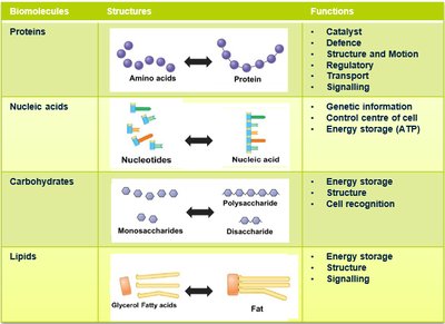
Overview of Biomolecules
Biomolecules are essential organic compounds that form the basis of life. The four major classes—proteins, nucleic acids, carbohydrates, and lipids—each have distinct structures and functions in the human body.
Biomolecules | Structures | Functions |
|---|---|---|
Proteins | Amino acids → Protein |
|
Nucleic acids | Nucleotides → Nucleic acid |
|
Carbohydrates | Monosaccharide → Disaccharide/Polysaccharide |
|
Lipids | Glycerol + Fatty acids → Fat |
|
Structure and Properties of Amino Acids
Basic Structure
Amino acids are the building blocks of proteins. Each amino acid contains a central (alpha) carbon atom bonded to four different groups: an amino group (NH2), a carboxyl group (COOH), a hydrogen atom, and a variable R-side chain.
Core elements: Carbon (C), hydrogen (H), oxygen (O), nitrogen (N)
R-side chain: Determines the unique properties of each amino acid
Stereochemistry
Amino acids (except glycine) are chiral and exist as L- and D- isomers. Only L-amino acids are incorporated into proteins in humans.
Classification by R-side Chains
The R-side chains of amino acids are grouped based on their chemical properties, which influence protein structure and function:
Small – hydrophobic
Branched-chain – hydrophobic
Hydroxyl – hydrogen bond
Sulfur – disulfide bond
Aromatic – hydrophobic
Acidic – H+ donor
Basic – H+ acceptor
Cyclic – proline only; rigidity creates bends in protein structure
Zwitterions and Acid-Base Properties
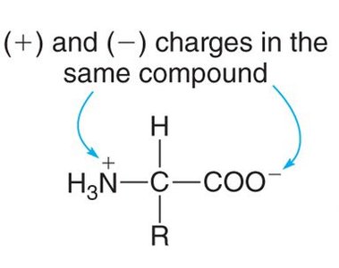
Amino acids can exist as zwitterions, molecules with both positive and negative charges but an overall neutral charge. Their charge state depends on the pH relative to their isoelectric point (pI):
At pH = pI: Zwitterion form (no net charge)
At pH < pI: Net positive charge (protonated carboxyl group)
At pH > pI: Net negative charge (deprotonated amino group)
Buffering Capacity
Amino acids act as buffers, resisting changes in pH by accepting or donating protons through their amino and carboxyl groups.
Peptide Bond Formation and Protein Structure
Peptide Bond Formation
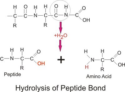
Two amino acids are joined by a peptide bond (amide bond) in a condensation reaction, releasing water (H2O).
N-terminus: Free amino group at the start of the chain
C-terminus: Free carboxyl group at the end of the chain
Levels of Protein Structure
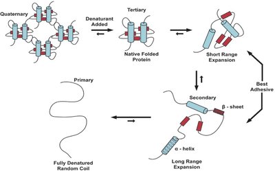
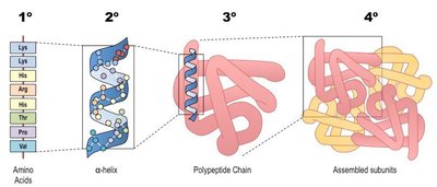
Proteins have four levels of structure, each contributing to their final shape and function:
Primary (1°) Structure: Linear sequence of amino acids joined by peptide bonds
Secondary (2°) Structure: Local folding into α-helices and β-pleated sheets, stabilized by hydrogen bonds
Tertiary (3°) Structure: Overall 3D shape of a single polypeptide, stabilized by interactions between R-groups (hydrogen bonds, ionic bonds, hydrophobic interactions, disulfide bonds)
Quaternary (4°) Structure: Assembly of multiple polypeptide subunits into a functional protein complex
Oligopeptides
Oligopeptides are short chains of amino acids (typically <20 residues) that do not fold into higher-order structures. Many peptide hormones are oligopeptides.
Protein Denaturation and Hydrolysis
Denaturation
Denaturation is the process by which proteins lose their secondary, tertiary, or quaternary structure due to disruption of non-covalent interactions and disulfide bonds. Common causes include heat, acids, bases, oxidizing/reducing agents, and heavy metals. The primary structure (peptide bonds) remains intact.
Hydrolysis
Hydrolysis is the cleavage of peptide bonds by the addition of water, breaking proteins down into individual amino acids. This process is catalyzed by enzymes during digestion.
Structure-Function Relationship in Proteins
Major Functions of Proteins
Catalysis (enzymes)
Defence (immunoglobulins)
Structure and motion (keratin, myosin)
Regulation (transcription factors like p53)
Transport (haemoglobin)
Signalling (insulin)
Examples of Structure-Function Relationships
Phenylalanine hydroxylase: Quaternary structure; single amino acid changes can cause phenylketonuria (PKU).
Immunoglobulin: Variable antigen-binding sites; sequence determines specificity and immune response.
Keratin: α-helices and disulfide bonds; structure determines properties of hair, nails, etc.
Myosin: Quaternary structure; mutations affect muscle contraction and can cause cardiomyopathy.
p53: DNA-binding domains; mutations disrupt gene regulation and can lead to cancer.
Haemoglobin: α and β subunits; mutations cause sickle-cell anaemia.
Insulin: Quaternary structure; mutations affect glucose regulation and can cause diabetes.
Clinical Relevance: Protein Disorders
Case Study: Rheumatic Heart Disease
Group A Streptococcal infections can lead to rheumatic heart disease due to molecular mimicry, where bacterial proteins resemble human heart proteins. The immune system attacks both, causing tissue damage. This highlights the importance of protein sequence and structure in immune recognition.
Case Study: Maple Syrup Urine Disease (MSUD)
MSUD is a metabolic disorder caused by a deficiency in the enzyme complex that breaks down branched-chain amino acids (leucine, isoleucine, valine). Accumulation of these amino acids and their toxic byproducts leads to neurological damage and can be fatal if untreated. Early detection via newborn screening is critical for management.
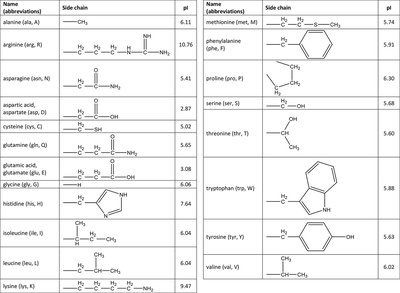
Summary Table: Amino Acids and Their Properties
Name (abbreviation) | Side chain | pI |
|---|---|---|
Alanine (Ala, A) | CH3 | 6.11 |
Arginine (Arg, R) | (CH2)3-NHC(NH)-NH2 | 10.76 |
Asparagine (Asn, N) | CH2-CONH2 | 5.41 |
Aspartic acid (Asp, D) | CH2-COOH | 2.87 |
Cysteine (Cys, C) | CH2-SH | 5.07 |
Glutamine (Gln, Q) | (CH2)2-CONH2 | 5.65 |
Glutamic acid (Glu, E) | (CH2)2-COOH | 3.22 |
Histidine (His, H) | CH2-imidazole | 7.64 |
Isoleucine (Ile, I) | CH(CH3)-CH2-CH3 | 6.04 |
Leucine (Leu, L) | CH2-CH(CH3)2 | 6.02 |
Lysine (Lys, K) | (CH2)4-NH2 | 9.47 |
Methionine (Met, M) | CH2-CH2-S-CH3 | 5.74 |
Phenylalanine (Phe, F) | CH2-phenyl | 5.48 |
Proline (Pro, P) | Pyrrolidine ring | 6.30 |
Serine (Ser, S) | CH2-OH | 5.68 |
Threonine (Thr, T) | CH(OH)-CH3 | 5.60 |
Tryptophan (Trp, W) | CH2-indole | 5.88 |
Tyrosine (Tyr, Y) | CH2-phenol | 5.66 |
Valine (Val, V) | CH(CH3)2 | 6.02 |
Key Equations
Peptide bond formation:
Hydrolysis of peptide bond:
Summary
Proteins are vital macromolecules with diverse structures and functions, determined by the sequence and properties of their constituent amino acids. Understanding protein structure, folding, and function is essential for appreciating their roles in health and disease, as illustrated by clinical cases such as PKU, MSUD, and rheumatic heart disease.
