뒤로
뒤로Amino Acids and Proteins: Structure, Properties, and Biological Roles
스터디 가이드 - 스마트 노트
 자료에 맞춘 맞춤형 노트, 핵심 정의, 예시, 맥락을 확장해 제공합니다.
WEEK 2 – AMINO ACIDS & PROTEINS
Topic I: Introduction to Biomolecules
Biomolecules are organic molecules that are essential for life and are found in all living organisms. They play critical roles in structure, function, and regulation of the body’s tissues and organs.
Four major biomolecules in the human body (from most to least abundant):
Proteins
Lipids
Carbohydrates
Nucleic acids
Structures and Functions of Biomolecules:
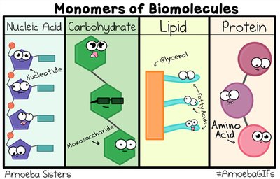
Proteins: Polymers of amino acids; function as enzymes, structural components, signaling molecules, and more.
Lipids: Hydrophobic molecules; serve as energy storage, membrane structure, and signaling molecules.
Carbohydrates: Polymers of monosaccharides; provide energy and structural support.
Nucleic acids: Polymers of nucleotides; store and transmit genetic information.
Common Properties Shared by All Biomolecules:
Carbon-based structure
Presence of functional groups
Ability to form polymers
Participation in noncovalent interactions
Importance of Noncovalent Interactions: Noncovalent interactions (hydrogen bonds, ionic bonds, van der Waals forces, and hydrophobic interactions) are crucial for the three-dimensional structure and biological activity of biomolecules. They allow for reversible interactions and dynamic biological processes.
Topic II: Biomolecules and Cells
Cells are the basic units of life, and their diversity in structure and function is due to the unique composition and arrangement of biomolecules.
Cellular Diversity: Different cell types arise due to differential gene expression and specialized biomolecule composition, allowing for a wide range of functions in the human body.
Biomolecule Composition in Cellular Structures:
Plasma membrane: Composed mainly of lipids (phospholipids, cholesterol), proteins, and carbohydrates.
Cytosol: Contains water, ions, proteins, carbohydrates, and small molecules.
Organelles: Each organelle has a unique composition; for example, mitochondria are rich in proteins and lipids, while the nucleus contains nucleic acids and proteins.
Cytoskeleton: Made up of protein filaments (actin, tubulin, intermediate filaments).
Universal Biomolecule: Proteins are present in the plasma membrane, cytosol, organelles, and cytoskeleton, reflecting their diverse roles in cellular structure and function.
Topic III: Basic Structures of Amino Acids
Amino acids are the building blocks of proteins. Each amino acid has a central carbon atom (α-carbon) bonded to four different groups.
Five Components of an Amino Acid:
Amino group (–NH2)
Carboxyl group (–COOH)
Hydrogen atom (–H)
R group (side chain, unique for each amino acid)
Central (α) carbon atom
Stereochemistry: Most amino acids (except glycine) are chiral and exist as L- and D-isomers. In proteins, only L-amino acids are found.
Eight Groups of R-Side Chains: Amino acids are classified based on the properties of their side chains: nonpolar aliphatic, aromatic, polar uncharged, positively charged (basic), negatively charged (acidic), sulfur-containing, hydroxyl-containing, and amide-containing.
Topic IV: Essential and Non-essential Amino Acids
Amino acids are categorized based on whether they can be synthesized by the human body.
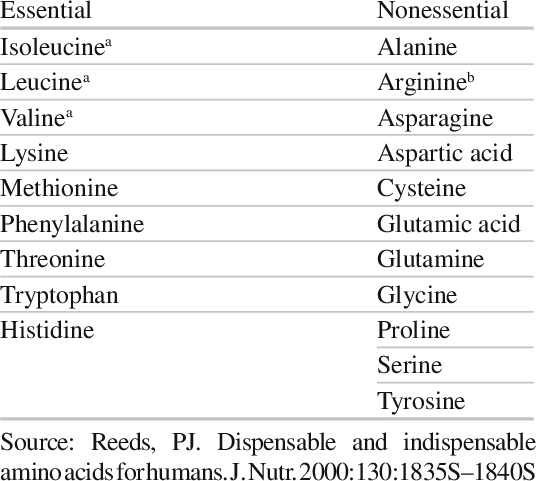
Essential Amino Acids: Cannot be synthesized by the body and must be obtained from the diet.
Non-essential Amino Acids: Can be synthesized by the body from metabolic intermediates.
Synthesis of Non-essential Amino Acids: These are produced via transamination and other metabolic pathways using intermediates from glycolysis and the citric acid cycle.
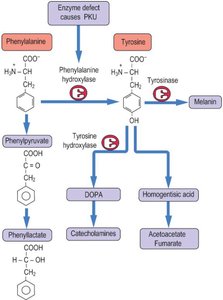
Phenylketonuria (PKU):
Causes: Deficiency of the enzyme phenylalanine hydroxylase.
Affected Amino Acids: Phenylalanine and tyrosine.
Consequences: Accumulation of phenylalanine leads to intellectual disability and other neurological problems.
Diagnosis: Newborn screening for elevated phenylalanine levels.
Treatment: Dietary restriction of phenylalanine.
Topic V: Amino Acids in Solutions
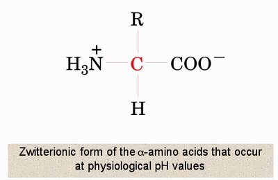
Amino acids can exist in different ionic forms depending on the pH of their environment, which affects their charge and buffering capacity.
Zwitterion: At physiological pH, amino acids exist as zwitterions, with a positively charged amino group and a negatively charged carboxyl group.
Isoelectric Point (pI): The pH at which an amino acid has no net charge (zwitterionic form).
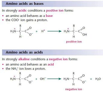
Charge States at Different pH Values:
At pH = pI: Amino acid is neutral (zwitterion).
At pH < pI: Amino acid is positively charged (amino group protonated).
At pH > pI: Amino acid is negatively charged (carboxyl group deprotonated).
Buffer: A buffer resists changes in pH upon addition of acid or base. Amino acids are excellent buffers due to their ionizable groups.
Topic VI: Peptide Bonds
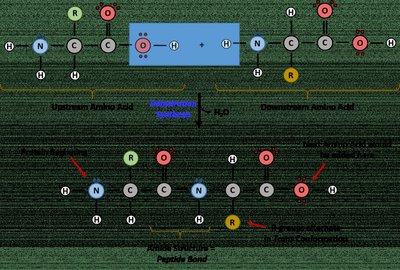
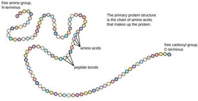
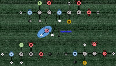
Peptide bonds link amino acids together to form peptides and proteins. This bond is formed through a condensation reaction between the amino group of one amino acid and the carboxyl group of another.
Formation of Peptide Bond: A covalent bond formed by the removal of a water molecule (dehydration synthesis) between the carboxyl group of one amino acid and the amino group of another.
Primary Structure of Proteins: The unique sequence of amino acids in a polypeptide chain.
Oligopeptide: A short chain of amino acids (typically 2–20 residues).
Topic VII: Protein Structures
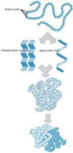
Proteins have four levels of structural organization, each contributing to their final shape and function.
Primary Structure: Linear sequence of amino acids.
Secondary Structure: Local folding into α-helices and β-pleated sheets stabilized by hydrogen bonds.
Tertiary Structure: Overall three-dimensional shape formed by interactions among R groups.
Quaternary Structure: Association of multiple polypeptide chains into a functional protein complex.
Topic VIII: Protein Denaturation & Hydrolysis
Proteins can lose their structure and function through denaturation or hydrolysis, which have distinct mechanisms and outcomes.
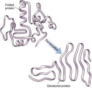
Denaturation: Disruption of secondary, tertiary, or quaternary structure without breaking peptide bonds. Caused by heat, pH changes, chemicals, or mechanical agitation. Results in loss of function.
Hydrolysis: Cleavage of peptide bonds, breaking the protein into individual amino acids. Catalyzed by enzymes or strong acids/bases.
Effects of Temperature and pH: Both can disrupt noncovalent interactions, leading to denaturation and loss of protein function, which can be detrimental to biological processes.
Topic IX: Structure-Function Relationships in Proteins
The structure of a protein determines its function. Alterations in structure can lead to loss or change of function, which is the basis for many diseases.
Six Major Functions of Proteins:
Catalysis (enzymes)
Defense (immunoglobulins)
Structure (keratin, collagen)
Movement (myosin, actin)
Regulation (transcription factors)
Transport (hemoglobin)
Signaling (insulin)
Importance of Structure for Function: The specific folding and arrangement of amino acids allow proteins to interact with other molecules and perform their biological roles. Mutations or denaturation can impair function.
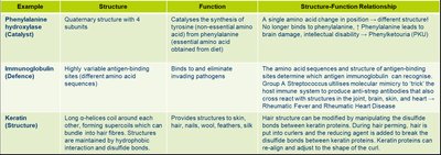
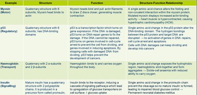
Example | Structure | Function | Structure-Function Relationship |
|---|---|---|---|
Phenylalanine hydroxylase (Catalytic) | Quaternary structure with 4 subunits | Catalyzes synthesis of tyrosine from phenylalanine | Mutation leads to PKU due to loss of function |
Immunoglobulin (Defense) | Highly variable antigen-binding sites | Binds and eliminates pathogens | Sequence variability allows recognition of diverse antigens |
Keratin (Structure) | Long α-helices coiled around each other | Provides structure to skin, hair, nails | Disulfide bonds stabilize structure |
Myosin (Motor) | Quaternary structure with subunits | Muscle contraction | Conformational changes generate force |
p53 (Regulatory) | Quaternary structure, DNA-binding domains | Regulates cell cycle | Mutations can lead to cancer |
Hemoglobin (Transport) | Quaternary structure, 2 α- and 2 β-subunits | Oxygen transport | Cooperative binding enhances function |
Insulin (Signaling) | Quaternary structure, 2 polypeptide chains | Regulates glucose uptake | Structure allows receptor binding |