 뒤로
뒤로Biological Membranes: Structure and Function – Biochemistry Study Guide
스터디 가이드 - 스마트 노트
 자료에 맞춘 맞춤형 노트, 핵심 정의, 예시, 맥락을 확장해 제공합니다.
Biological Membranes: Structure and Function
Overview of Membrane Structure and Function
Biological membranes are essential for cellular compartmentalization, selective transport, and signal transduction. They are composed of lipids, proteins, and carbohydrates, forming dynamic, asymmetric structures that regulate the flow of molecules and information.
Membrane lipids are synthesized in the smooth endoplasmic reticulum.
Membrane composition affects flexibility, permeability, and compartmentalization.
Functions: boundary definition, selective import/export, retention of metabolites, signal sensing, compartmentalization, and energy gradient generation.
Lipids: Structure and Properties
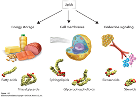
Lipids are a heterogeneous class of organic compounds, insoluble in water but soluble in organic solvents. Their amphipathic nature is crucial for membrane formation.
Amphipathic molecules: possess both polar (hydrophilic) and nonpolar (hydrophobic) regions.
Major types: fats, oils, phospholipids, sphingolipids, cholesterol.
Fatty Acids: Classification and Structure
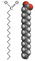
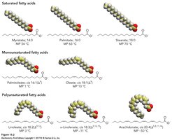
Fatty acids are carboxylic acids with hydrocarbon chains (4–36 carbons), forming the backbone of many lipids. They are classified by saturation and double bond configuration.
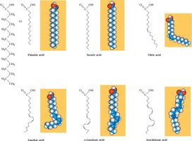
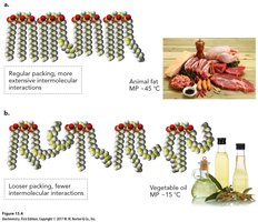
Saturated: no double bonds; pack tightly, higher melting points.
Unsaturated: one or more double bonds; cis isomers predominate, causing kinks and lower melting points.
Monounsaturated: one double bond.
Polyunsaturated: multiple double bonds; essential fatty acids (e.g., linoleate, α-linoleate, arachidonate) must be obtained from the diet.
Trans fatty acids: formed by hydrogenation, resemble saturated fatty acids, associated with cardiovascular disease.
Aggregation of Lipids in Water
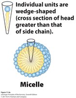
The amphipathic nature of lipids drives their aggregation into micelles, vesicles, and bilayers, which are fundamental to membrane structure.
Micelles: spherical structures with a single polar surface.
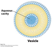
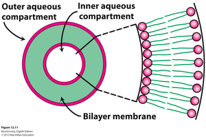
Vesicles (liposomes): bilayered spheres with an aqueous cavity, useful for drug delivery.
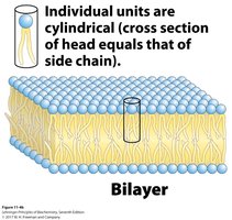
Bilayers: two sheets of lipids forming the basic structure of biological membranes.
Membrane Bilayer: Formation and Stabilization
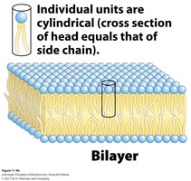
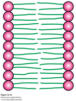
Membrane bilayers form when lipids with polar head groups and multiple tails are in aqueous solution. The hydrophobic effect and van der Waals interactions stabilize the bilayer.
Hydrophilic heads: interact with water.
Hydrophobic tails: interact with each other, forming the membrane's interior.
Stabilization: van der Waals interactions between tails.
Fluid Mosaic Model of Membranes
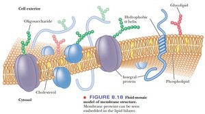
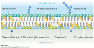
The fluid mosaic model describes membranes as dynamic, asymmetric structures with lipids and proteins. Lipids and proteins can diffuse laterally, and the composition varies between leaflets.
Asymmetry: different lipids and proteins on inner and outer leaflets.
Glycolipids: found on the outer leaflet, important for cell recognition.
Electrical polarization: membranes can be polarized.
Major Types of Membrane Lipids
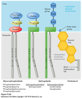
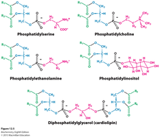
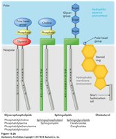
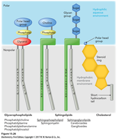
Membranes contain three major types of lipids: phospholipids (glycerophospholipids), sphingolipids, and cholesterol. These lipids can be diversified by modifying their backbone, fatty acids, and head groups.
Phospholipids: most abundant, form bilayers.
Sphingolipids: contain sphingosine, important in nerve cells.
Cholesterol: modulates fluidity and permeability.
Sphingolipids and Glycolipids
Sphingolipids are found in membranes, especially nerve cells. Glycolipids are carbohydrate-containing lipids derived from sphingosine, important for cell-cell recognition.
Sphingomyelin: enriched in nerve cells.
Cerebrosides: contain simple sugars.
Gangliosides: contain branched oligosaccharides.
Cholesterol: Structure and Function
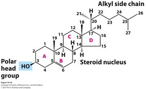
Cholesterol is a major component of eukaryotic membranes, affecting fluidity and permeability. It is absent from prokaryotic and fungal cells and serves as a precursor for steroid hormones and bile acids.
Structure: 27 carbons, four-ring steroid core, hydroxyl group interacts with phospholipid heads.
Function: modulates membrane fluidity, precursor for hormones.
Membrane Lipids Are Amphipathic
Membrane lipids have both hydrophilic and hydrophobic portions, orienting themselves to form bilayers with hydrophilic heads facing water and hydrophobic tails facing inward.
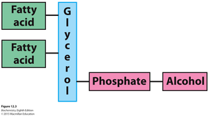
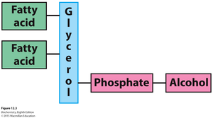
Hydrophilic head: phosphate and alcohol components.
Hydrophobic tail: fatty acid chains.
Membrane Fluidity and Dynamics
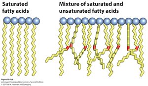
Membrane fluidity is controlled by fatty acid composition and cholesterol content. Saturated fatty acids increase rigidity, while unsaturated fatty acids and cholesterol increase fluidity.
Saturated fatty acids: linear, rigid packing.
Unsaturated fatty acids: cis double bonds cause disorder and fluidity.
Cholesterol: disrupts tight packing, modulates fluidity.
Membrane Dynamics: Lateral and Transverse Diffusion
Lipids and proteins can diffuse laterally within the bilayer, but transverse diffusion (flip-flop) is rare and often catalyzed by enzymes called flippases.
Lateral diffusion: rapid movement within the same leaflet.
Transverse diffusion: slow, requires flippases, sometimes ATP-dependent.
Membrane Rafts
Lipid rafts are microdomains within membranes, enriched in cholesterol and sphingolipids, important for cell signaling and protein segregation.
Rafts: clusters of proteins and lipids, facilitate signaling.
Membrane Proteins: Types and Functions
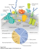
Membrane proteins are integral to membrane function, serving as receptors, channels, pumps, enzymes, and anchors. They are classified as integral, peripheral, or amphitropic.
Integral proteins: embedded in the membrane, often span the bilayer.
Peripheral proteins: loosely associated, can be removed easily.
Amphitropic proteins: conditionally attached, regulated by lipid or carbohydrate interactions.
Amino Acid Residues in Membrane Proteins
The placement of amino acid residues in membrane proteins is critical for their function. Hydrophobic residues are found in transmembrane segments, while polar and charged residues are located in aqueous domains.
Hydrophobic residues: interact with lipid tails.
Tyr and Trp: cluster at nonpolar/polar interfaces.
Charged residues: found in aqueous domains.
Membrane Permeability and Transport
Membranes are highly impermeable to ions and most polar molecules. Transport across membranes is facilitated by proteins and can be passive or active.
Passive transport: simple diffusion, facilitated diffusion (no energy required).
Active transport: requires energy, moves substances against gradients.
Transporters: integral proteins, serve as pores and channels.
Passive Transport: Simple and Facilitated Diffusion
Passive transport moves solutes down their concentration or electrochemical gradient. Facilitated diffusion uses carrier proteins or channels for specific substrates.
Simple diffusion: O2, CO2, fat-soluble vitamins.
Facilitated diffusion: carbohydrates, amino acids, ions.
Ion Channels and Porins
Ion channels and porins facilitate rapid, selective transport of ions and water across membranes. Ion channels are gated and regulated by biological signals.
Ion channels: specificity for ions, not saturable, gated.
Porins: form hydrophilic channels for water (e.g., aquaporin).
Active Transport: Primary and Secondary
Active transport moves substances against their concentration gradient, requiring energy. Primary active transport uses ATP hydrolysis, while secondary active transport uses ion gradients.
Primary: Na+–K+ pump.
Secondary: Na+-glucose symporter.
Summary Table: Major Membrane Lipids
Lipid Type | Structure | Function |
|---|---|---|
Phospholipids | Glycerol backbone, fatty acids, phosphate, alcohol | Bilayer formation, compartmentalization |
Sphingolipids | Sphingosine backbone, fatty acid, head group | Cell recognition, nerve cell membranes |
Cholesterol | Four-ring steroid core, hydroxyl group | Fluidity modulation, precursor for hormones |
Membrane Structure and Function Summary
Membranes are sheetlike structures, two molecules thick, forming boundaries of cells and organelles. They consist of lipids and proteins, with some proteins containing carbohydrate moieties. Membrane lipids are amphipathic, and associated proteins serve as pumps, channels, receptors, and enzymes. Membranes are asymmetric, fluid, and control the flow of solutes and ions.
