 뒤로
뒤로Biological Membranes: Structure and Function – Study Notes
스터디 가이드 - 스마트 노트
 자료에 맞춘 맞춤형 노트, 핵심 정의, 예시, 맥락을 확장해 제공합니다.
Biological Membranes: Structure and Function
Introduction to Biological Membranes
Biological membranes are essential structures that define the boundaries of cells and organelles, enabling compartmentalization and regulation of biochemical processes. They are primarily composed of lipids and proteins, with some carbohydrates, and exhibit unique properties such as fluidity, asymmetry, and selective permeability.
Lipids in Biological Membranes
Definition and Properties of Lipids
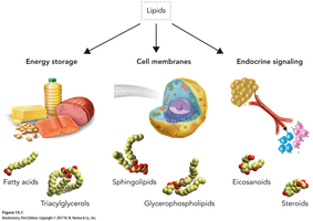
Lipids are a heterogeneous class of naturally occurring organic compounds, classified by their solubility in organic solvents and insolubility in water.
They are amphipathic, meaning they possess both hydrophilic (polar) and hydrophobic (nonpolar) regions.
Major examples include fats, oils, phospholipids, sphingolipids, and cholesterol.
Types of Lipids
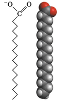
Fatty acids: Carboxylic acids with hydrocarbon chains (4–36 carbons), usually unbranched and with an even number of carbons.
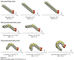
Saturated fatty acids: No double bonds; straight chains.
Unsaturated fatty acids: One or more double bonds; can be monounsaturated (one double bond) or polyunsaturated (multiple double bonds).
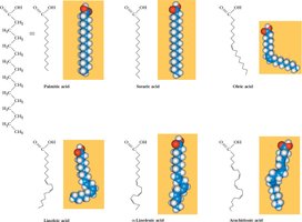
Fatty acids are named with the suffix “-ate” at physiological pH (e.g., palmitate, oleate).
Common Fatty Acids: Structures and Nomenclature
Essential fatty acids (e.g., linoleate, α-linolenate, arachidonate) must be obtained from the diet.
Polyunsaturated fatty acids are identified by the position of the double bond from the omega (ω) carbon.
Physical Properties: Saturated vs. Unsaturated Fatty Acids
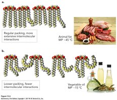
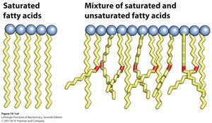
Cis double bonds introduce kinks, preventing tight packing and lowering melting points.
Trans fatty acids (rare in nature, formed by hydrogenation) resemble saturated fatty acids in shape and are associated with health risks.
Greater unsaturation leads to lower melting points.
Melting Point and Double Bonds
Saturated fatty acids pack tightly, resulting in higher melting points (e.g., animal fats).
Unsaturated fatty acids pack loosely, resulting in lower melting points (e.g., vegetable oils).
Membrane Structure: Lipid Aggregates
Amphipathic Lipids and Aggregate Structures
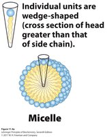
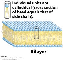
Amphipathic lipids spontaneously form organized structures in water due to the hydrophobic effect. The three major structures are:
Micelles: Spherical structures with a single layer of lipids, polar heads outward, hydrophobic tails inward.
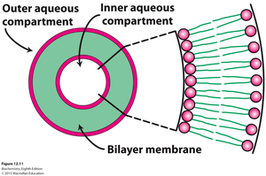
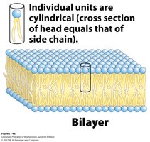
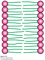
Bilayers: Two layers of lipids with hydrophobic tails facing inward and hydrophilic heads facing the aqueous environment.
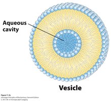
Vesicles (Liposomes): Spherical bilayers enclosing an aqueous cavity, useful for drug delivery.
Membrane Bilayer
Composed of two lipid sheets stabilized by van der Waals interactions between hydrophobic tails.
Hydrophilic head groups interact with water on both sides.
Major lipid types: glycerophospholipids and sphingolipids.
Membrane Composition and Function
Overview and Function of Membranes
Membranes define cell boundaries, allow selective import/export, retain metabolites, and sense external signals.
They provide compartmentalization, separate energy-producing and energy-consuming reactions, and support ATP synthesis via proton gradients.
Fluid Mosaic Model
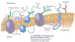
Membranes are dynamic, asymmetric structures composed of lipids, proteins, and carbohydrates.
Lipids and proteins can move laterally within the bilayer.
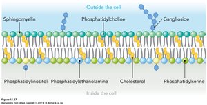
Glycolipids are typically found on the outer leaflet.
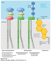
Major Types of Membrane Lipids
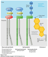
Phospholipids (including glycerophospholipids and sphingolipids)
Cholesterol
Lipids can be diversified by modifying their backbone, fatty acids, and head groups.
Phospholipids and Glycerophospholipids
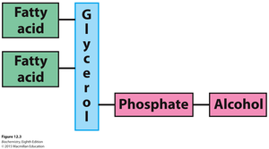
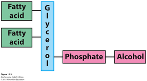
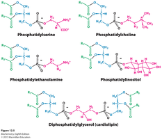
Phospholipids are constructed from fatty acids, glycerol, phosphate, and an alcohol group.
Glycerophospholipids are the most abundant membrane lipids; their head groups determine membrane surface properties.
Sphingolipids
Contain a sphingosine backbone linked to a fatty acid.
Include sphingomyelin (with phosphorylcholine), cerebrosides (with simple sugars), and gangliosides (with oligosaccharides).
Glycolipids play roles in cell-cell recognition.
Cholesterol
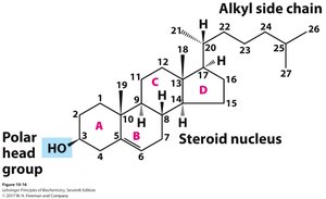
Comprises 25–40% of plasma membrane lipids in animals.
Rigid four-ring structure; hydroxyl group interacts with phospholipid head groups.
Modulates membrane fluidity and permeability; precursor for steroid hormones and bile acids.
Amphipathic Nature of Membrane Lipids
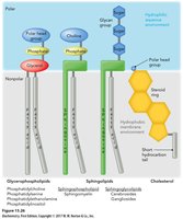
Membrane lipids have both hydrophilic (polar head) and hydrophobic (fatty acid tail) regions.
This property drives the formation of bilayers and other structures in aqueous environments.
Membrane Proteins
Types of Membrane Proteins
Integral (intrinsic) proteins: Embedded within the membrane, often spanning the bilayer.
Peripheral (extrinsic) proteins: Loosely associated with the membrane via electrostatic interactions or lipid anchors.
Amphitropic proteins: Can reversibly associate with membranes, often regulated by covalent modification.
Integral Membrane Proteins
Monotopic: Interact with one leaflet; Polytopic: Span both leaflets.
Hydrophobic stretches interact with lipid tails; can be removed by detergents.
Example: Bacteriorhodopsin (polytopic, contains membrane-spanning α-helices).
Beta-Barrel Membrane Proteins
Composed of β-sheets forming a hollow cylinder (pore or channel).
Hydrophobic exterior interacts with membrane; polar interior allows passage of molecules.
Amino Acid Distribution in Membrane Proteins
Transmembrane segments are rich in hydrophobic residues.
Tyr and Trp cluster at the interface; charged residues are found in aqueous regions.
Peripheral and Amphitropic Proteins
Peripheral proteins associate via electrostatic interactions or covalent lipid anchors (e.g., GPI anchors).
Amphitropic proteins can reversibly associate with membranes, regulated by lipidation or other modifications.
Membrane Properties and Dynamics
Lipid Bilayer Properties
Held together by noncovalent interactions (van der Waals, hydrophobic effect).
Inner and outer leaflets have different lipid compositions; outer layer is often more positively charged.
Membrane Fluidity
Controlled by fatty acid composition and cholesterol content.
Saturated fatty acids increase rigidity; unsaturated fatty acids and cholesterol increase fluidity.
Membrane Dynamics: Lateral and Transverse Diffusion
Lipids and proteins can move laterally within the bilayer (lateral diffusion).
Transverse (flip-flop) diffusion is rare and catalyzed by enzymes called flippases, which may require ATP.
Membrane Rafts
Microdomains enriched in cholesterol, sphingolipids, and certain proteins.
Serve as platforms for cell signaling and protein segregation.
Membrane Transport
Membrane Permeability
Lipid bilayers are highly impermeable to ions and most polar molecules; water can diffuse slowly.
Membrane proteins facilitate the transport of molecules and information.
Types of Membrane Transport
Passive (simple) diffusion: Movement down a concentration gradient without energy input (e.g., O2, CO2).
Facilitated diffusion: Movement via specific transporters or channels, still down a gradient and without energy input.
Active transport: Movement against a gradient, requiring energy (e.g., ATP hydrolysis).
Passive Transport and Facilitated Diffusion
Facilitated diffusion allows polar molecules and ions to cross membranes via specific proteins (e.g., glucose transporter, ion channels).
Transporters reduce the activation energy for transmembrane movement by providing a hydrophilic pathway.
Ion Channels and Aquaporins
Ion channels allow rapid, selective movement of ions; regulated by gates.
Aquaporins facilitate bulk water transport across membranes.
Ionophores
Small molecules (often antibiotics) that shuttle ions across membranes, disrupting gradients and cellular processes.
Transport Systems: Uniport, Symport, Antiport
Uniport: Transports one type of molecule.
Symport: Transports two molecules in the same direction.
Antiport: Transports two molecules in opposite directions.
Glucose Transport (GLUT1)
GLUT1 alternates between two conformations to transport glucose across the membrane down its concentration gradient.
Bicarbonate Transporter (Antiporter)
Exchanges HCO3- and Cl- across the membrane, maintaining electrochemical balance.
Active Transport
Primary active transport: Directly uses ATP hydrolysis (e.g., Na+/K+ pump).
Secondary active transport: Uses ion gradients established by primary transport (e.g., Na+-glucose symporter).
Sodium–Potassium Ion Pump Mechanism
Transports 3 Na+ out and 2 K+ in per ATP hydrolyzed, maintaining gradients essential for cell function.
Efficient Glucose Import
Na+-glucose symporter (apical side) and glucose uniporter (basolateral side) work together to absorb glucose from the intestine into the blood.
Summary Table: Major Membrane Lipids
Lipid Type | Backbone | Head Group | Key Features |
|---|---|---|---|
Glycerophospholipids | Glycerol | Phosphate + Alcohol | Major membrane lipid; amphipathic |
Sphingolipids | Sphingosine | Phosphate or Sugar | Cell recognition, nerve tissue |
Cholesterol | Steroid nucleus | Hydroxyl group | Modulates fluidity, precursor for hormones |
Key Equations
Passive diffusion rate:
Electrochemical potential:
Conclusion
Biological membranes are complex, dynamic structures essential for cellular compartmentalization, signaling, and transport. Their unique composition of lipids and proteins underlies their diverse functions and adaptability in various physiological contexts.
