뒤로
뒤로Biosignaling: Mechanisms and Pathways in Cellular Communication
스터디 가이드 - 스마트 노트
 자료에 맞춘 맞춤형 노트, 핵심 정의, 예시, 맥락을 확장해 제공합니다.
Biosignaling
Introduction to Biosignaling
Biosignaling refers to the complex processes by which cells communicate with each other and respond to external stimuli. This communication is essential for maintaining homeostasis, regulating metabolism, and coordinating cellular functions. The process involves chemical messengers, receptors, and intricate signaling pathways that translate extracellular signals into specific cellular responses.
Objectives of Biosignaling Study
Terminology: Understanding and correctly using biochemistry-related terms.
Membrane Components: Identifying major membrane components and their roles in compartmentation, membrane potential, transport, and anchoring.
Receptor-Ligand Interactions: Describing properties and roles in signal transduction and disease.
Types of Intercellular Communication: Comparing five types of chemical mediator-based communication.
Signaling Processes: Contrasting intracellular and cell-surface receptor signaling.
GPCR Signaling: Identifying primary and secondary messengers.
Enzyme-Coupled and Ion-Channel Receptors: Describing their signaling processes.
Signal Termination: Explaining methods of signal cessation.
Cellular Communication
Mechanisms of Cell-to-Cell Communication
Cells communicate through proteins that form pathways to respond to extracellular signals. Receptors on the cell surface or within the cell sense these signals and initiate a cascade of events leading to a cellular response. These receptors serve as the critical link between external signals and internal cellular responses. 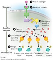
Signal Transduction Pathway
Overview of Signal Transduction
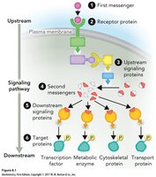
Signal transduction is the biochemical mechanism by which extracellular signals are transmitted across the cell membrane. A ligand (first messenger) binds to a receptor protein, triggering a cellular response.
Agonist: A ligand that initiates a biological response upon binding to a receptor.
Antagonist: A ligand that binds to a receptor but does not elicit a response.
Signal transduction pathways often result in covalent or noncovalent modification of intracellular target proteins.
General Features of Chemical Messengers
Properties and Specificity
Chemical messengers (ligands) elicit responses in target cells. The specificity of the response depends on the type and location of the receptor and the specific messenger. Target cells possess receptors for the messengers they can respond to. Activation of a receptor may involve:
Covalent protein modifications
Protein conformational changes
Alteration in gene expression rates
Intercellular Communication by Chemical Mediators
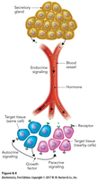
Types of Intercellular Communication
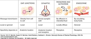
Cells communicate via several mechanisms, each with distinct properties and specificity.
Type | Message Transmission | Local or General | Specificity Depends On |
|---|---|---|---|
Gap Junctions | Directly from cell to cell | Local | Anatomic location |
Synaptic | Across synaptic cleft | Local | Anatomic location and receptors |
Paracrine and Autocrine | By diffusion in interstitial fluid | Locally diffuse | Receptors |
Endocrine | By circulating body fluids | General | Receptors |
General Steps of Chemical Messenger Action
Sequence of Events
The chemical messenger (ligand) is secreted from a specific cell in response to a stimulus.
The messenger diffuses or is transported through blood or extracellular fluid to the target cell.
Hydrophobic messengers diffuse across the plasma membrane and bind to intracellular receptors, eliciting a response.
Receptors on the target cell membrane specifically bind the messenger.
Binding of the messenger to the receptor elicits a response or the signal is terminated.
Types of Primary Messengers
Representative First Messengers
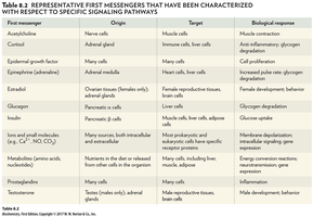
Primary messengers are diverse and include neurotransmitters, hormones, and growth factors.
First Messenger | Origin | Target | Biological Response |
|---|---|---|---|
Acetylcholine | Neurons | Muscle cells | Muscle contraction |
Cortisol | Adrenal gland | Muscle cells, liver cells | Anti-inflammatory, glycogen degradation |
Epidermal growth factor | Many cells | Many cells | Cell proliferation |
Epinephrine | Adrenal gland | Heart cells, liver cells | Increased pulse rate, glycogen degradation |
Glucagon | Pancreas | Muscle cells, liver cells | Glucose uptake |
Metabolites (e.g., Ca2+, NO, CO2) | Many sources | Many cells | Metabolic regulation |
Second Messengers
Role and Examples
Second messengers are small, non-protein intracellular molecules that amplify receptor-generated signals. Common examples include:
Cyclic AMP (cAMP)
Cyclic GMP (cGMP)
Diacylglycerol (DAG)
Inositol-1,4,5-triphosphate (IP3)
Ca2+
These molecules amplify the signal initiated by a primary messenger, leading to a robust cellular response.
Cell Surface vs. Intracellular Receptors
Comparison of Receptor Types
Cell-Surface Receptors: Bind large proteins or polar molecules (e.g., peptide hormones, cytokines, catecholamines) that cannot cross the membrane rapidly.
Intracellular Receptors: Bind hydrophobic messengers (e.g., steroid hormones) that diffuse through the membrane. These receptors act as gene-specific transcription factors, regulating gene expression.
Nuclear Receptor Signaling
Mechanism of Nuclear Receptor Action
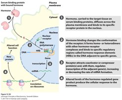
Nuclear receptors function as transcription factors that regulate gene expression. Steroid hormones, being lipids, enter cells by simple diffusion, while thyroid hormones use facilitated diffusion. Lipophilic hormones are transported in the blood bound to serum albumin, steroid hormone-binding globulin (SHBG), or thyroid hormone-binding globulin (TBG). These hormones can activate or inhibit transcription of specific genes. 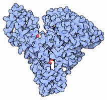
Direct Regulation of Transcription by Hormones
Hormone Response Elements (HRE)
Ligand binding causes a conformational change in the nuclear receptor, allowing it to bind DNA. Activated receptors bind to hormone response elements (HRE), which are short, specific DNA sequences in promoters of hormone-responsive genes. This process can increase or decrease protein production.
Example: Cortisol Binding to the Glucocorticoid Receptor
Mechanism of Action
The glucocorticoid receptor (GR) is located in the cytosol, bound to heat shock proteins (HSP). When cortisol binds GR, it dissociates from HSP, exposing a nuclear localization signal (NSL). GR homodimerizes and translocates to the nucleus, binding to HREs in DNA to regulate transcription of genes involved in metabolism.
Cell-Surface Receptors
Structure and Function
All membrane receptors share common features:
Extracellular domain for ligand binding
One or more membrane-spanning α-helices
Intracellular domain for signal transduction
Ligand binding to the extracellular domain causes a conformational change, activating the intracellular domain and initiating a signal transduction pathway. Effects include rapid changes in ion concentration, enzyme activation/inhibition, and changes in gene expression.
Common Membrane Receptors
Types of Membrane Receptors
Ion channel receptors
Receptors that are kinases or activate kinases
Receptors that work through second messengers
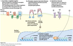
G Protein−Coupled Receptor (GPCR) Signaling
Structure and Function of GPCRs
GPCRs are α-helical integral membrane proteins that traverse the membrane seven times. Humans have over 600 GPCR genes, many involved in sensory responses. 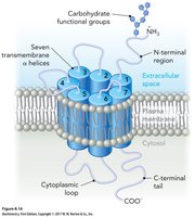
GPCRs Activate Heterotrimeric G Proteins
Mechanism of G Protein Activation
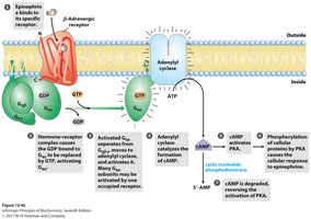
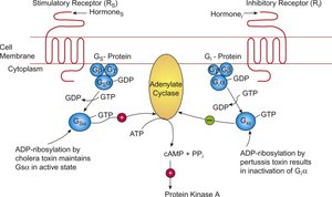
G-proteins are heterotrimeric (αβγ) membrane-associated proteins that bind GTP. The G protein complex with GDP is inactive. Upon signal transmission, GPCR undergoes a conformational change, leading to GDP-GTP exchange on the α subunit, which then dissociates from βγ. 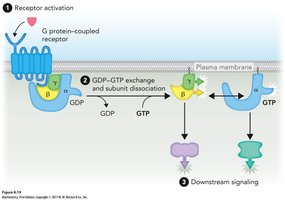
Downstream Signaling of GPCR
Types of G Proteins and Their Effects
Gsα (stimulatory): Activates adenylate cyclase; second messenger is cAMP.
Giα (inhibitory): Inhibits adenylate cyclase.
Gqα (stimulatory): Stimulates phospholipase C (PLC); second messengers are IP3, DAG, and Ca2+.
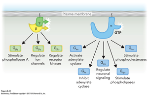
Sensing the Epinephrine Signal via GPCR
Epinephrine Signaling
Epinephrine, produced by adrenal glands, mediates the stress response by mobilizing energy. Its effects vary by tissue:
Muscle/liver: Glycogen breakdown
Adipose: Lipid hydrolysis
Heart: Increased heart rate and contractility
Signal Amplification by cAMP
Amplification Mechanism
Second messengers like cAMP amplify the signal initiated by a primary messenger. Receptor activation of adenylate cyclase generates cAMP, which activates Protein Kinase A (PKA). PKA phosphorylates downstream enzymes, leading to the release of thousands of glucose molecules. 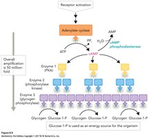
Self-Inactivation in G-protein Signaling
Termination of Signal
Epinephrine is a short-acting signal. The α subunit of G protein (Gα) is a GTPase, hydrolyzing GTP to GDP, restoring the inactive form and re-associating with βγ. 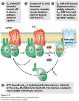
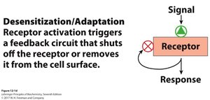
Additional Forms of Inactivation of Gαs
Inhibitory G Proteins and Phosphodiesterase
Giα inhibits adenylate cyclase when bound to GTP.
Cyclic nucleotide phosphodiesterase cleaves cAMP into AMP, terminating the signal.
GPCRs Can Use Other Secondary Messenger Molecules
Phospholipase C Pathway
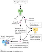
Epinephrine binding to the α1-adrenergic receptor activates Gqα, stimulating PLC. PLC cleaves PIP2 into IP3 and DAG, which activate different signaling pathways. 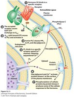
Function and Properties of Second Messengers
PIP2, IP3, DAG, and Ca2+
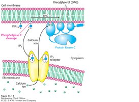
PIP2 is cleaved by PLC into IP3 and DAG.
IP3 binds to ligand-gated ion channels on the ER, causing Ca2+ release.
DAG remains in the membrane and activates PKC.
Secondary Messengers Can Activate Other Secondary Messenger Molecules
Calcium Signaling
GPCR activation of IP3 can open ligand-gated ion channels, releasing Ca2+ which activates calcium-sensing proteins like PKC or calmodulin, leading to phosphorylation of target enzymes. 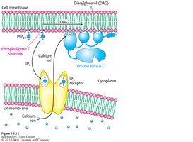
Second Messengers: PLC Pathway
PLC and Downstream Effects
PLC hydrolyzes PIP2 into IP3 and DAG.
DAG activates PKC.
IP3 activates Ca2+ channels in the ER, increasing cytoplasmic Ca2+.
Ca2+ activates calmodulin, which can phosphorylate target enzymes.
Signaling Pathways that Affect Transcription
Pathways to Transcriptional Regulation
Three distinct pathways for primary messengers to alter transcription:
Steroid hormone receptors (e.g., glucocorticoid receptor)
Activation of protein kinases in cytosol that move to the nucleus (e.g., MAPK cascade)
Activation of latent transcription factors in cytosol (e.g., JAK-STAT, NF-κB)
Enzyme-Coupled Receptors: Kinases
Kinase Receptor Mechanism
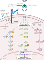
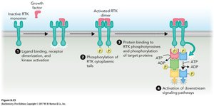
Kinases transfer phosphate groups from ATP to specific amino acid residues (Ser, Thr, Tyr) on target proteins. Ligand binding activates the intracellular kinase domain, leading to autophosphorylation or phosphorylation of associated proteins. 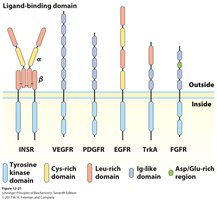
Growth Factors and Tyrosine Kinase Receptors
Regulation of Cell Growth
Growth factors regulate gene expression affecting growth, differentiation, migration, and cell functions. The pathway leads to activation of MAPK, phosphorylation of nuclear transcription factors Jun and Fos, and activity of E2F, promoting synthesis of enzymes essential for DNA synthesis. 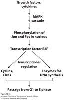
JAK-STAT Pathway Inhibition
Pharmacological Inhibition
Toficitinib (Xeljanz) is a pan Janus kinase (JAK) inhibitor that interferes with the JAK-STAT pathway, preventing phosphorylation and dimerization of STAT, thus blocking cytokine gene transcription. 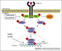
Ion-Channel Receptors
Regulation of Ion Transport
Ion-channel receptors regulate the transport of ions (Cl-, K+, Na+, Ca2+) across cell membranes. They respond to changes in membrane potential or ligand binding and play key roles in the nervous system. Examples include voltage-gated sodium channels, nicotinic acetylcholine receptor, ionotropic glutamate receptor, and gamma aminobutyric acid receptor A.
Membranes Are Electrically Polarized
Membrane Potential
The inside of the cell is typically negatively charged compared to the outside (Vm –50 to –70 mV). This polarization is due to asymmetric transport of cations by Na+K+ ATPase, which moves 3 Na+ out and 2 K+ in. 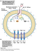
Voltage-Gated and Ligand-Gated Ion Channels in Neuronal Signaling
Neuronal Signal Propagation
Signals within neurons propagate as electrical impulses, involving opening of voltage-gated Na+ channels. Opening of voltage-gated Ca2+ channels at the axon end triggers release of neurotransmitter acetylcholine, which opens ligand-gated ion channels on the receiving cell, continuing the action potential. 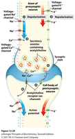
Nicotinic Acetylcholine Receptor
Mechanism of Action
Acetylcholine (ACh) acts on nicotinic ACh receptors on muscle cells. Neurotransmitters are secreted in response to an action potential, causing voltage changes across the membrane. ACh diffuses across the synapse, binds to receptors, and elicits a response. Before release, ACh is sequestered in vesicles near the presynaptic membrane. Voltage-gated Ca2+ channels open upon action potential arrival, triggering vesicle fusion and ACh release. ACh binding causes conformational change in the receptor, opening ligand-gated Na+/K+ channels, leading to muscle contraction. Acetylcholinesterase breaks down ACh to terminate the signal.
Signal Termination
Methods of Signal Termination
Signals for metabolic processes or nerve impulses must be rapidly terminated when the messenger is no longer produced. Proliferation signals take longer to terminate. Failure in signal termination can lead to disease.
Removal of stimulus or diffusion away from receptor
Phosphatases remove phosphate groups
GTPases break down G proteins
Phosphodiesterases degrade cAMP
Summary
Key Concepts
Cells communicate via chemical messengers binding to plasma membrane receptors, initiating intracellular changes.
Receptor families include ion channels, GPCRs, and enzyme-linked receptors.
Activation of cytosolic steroid receptors leads to changes in membrane potential, G-protein activation, second messenger increase, or transcription initiation.
Second messengers undergo rapid concentration changes following primary messenger binding. Common examples: Ca2+, cAMP, cGMP, IP3, DAG.